IS Families/IS6 family
This chapter has appeared in a modified form as: The IS6 family, a clinically important group of insertion sequences including IS26. Alessandro Varani, Susu He, Patricia Siguier, Karen Ross and Michael Chandler.Mobile DNA (2021) 12:11 doi:10.1186/s13100-021-00239-x [1]
Contents
- 1 General
- 2 Distribution and Phylogenetic Transposase Tree
- 3 Terminal Inverted Repeats.
- 4 Genomic Impact and Clinical Importance
- 5 Organization
- 6 Mechanism: the state of play
- 7 Cointegrate formation
- 8 Circular transposon molecules: translocatable units (TU)
- 9 Targeted transposition.
- 10 A Model for Targeted Integration
- 11 Emerging Biochemistry: Transposase binding.
- 12 Conclusions and Future Directions
- 13 Acknowledgements
- 14 Bibliography
General
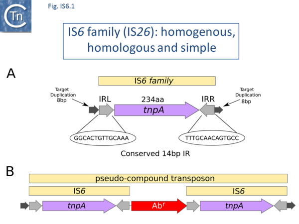
There are at present nearly 160 family members in ISfinder from nearly 80 bacterial and archaeal species but this represents only a fraction of those present in the public databases. The family was named[2] after the directly repeated insertion sequences in transposon Tn6 [3] to standardize the various names that had been attributed to identical elements (e.g. IS15Δ/IS15D, IS26, IS46, IS140, IS160, IS176) [4][5][6][7][8][9][10][11][12][13][14][15], including one isolate, IS15, corresponding to an insertion of one iso-IS6 (IS15Δ/IS15D, note that IS15Δ is sometimes referred to as IS15D, IS15-Δ or IS15-delta) into another [5][6] (Note that compared to IS26, IS15D derivatives, have one or two base pair changes). More recently there has been some attempt to rename the family as the IS26 family (see [16]), presumably because of accumulating experimental data from IS26 itself and the importance of this IS in accumulation and transmission of multiple anti biotic resistance, although this might potentially introduce confusion in the literature. IS6 family members have a simple organization (Fig. IS6.1) and generate 8bp direct target repeats on insertion.
This family is very homogenous with an average length of about 800 bp and highly conserved short, generally perfect, IRs (Fig. IS6.1 and Fig. IS6.2). There are two examples of MITES (Miniature Inverted repeat Transposable Elements composed of both IS ends and no intervening orfs; [17]of 227 and 336 bp), 7 members between 1230 and 1460 bp and three members between 1710 and 1760 bp. One member, IS15, of 1648 bp represents and insertion of one IS into another [4][6]. Many are found as part of compound transposons (called pseudo-compound transposons [2] described below) invariably as flanking direct repeats (Fig. IS6.1) a consequence of their transposition mechanism [7][9][13][14][18][19][20][21][22][23][24][25][26][27][28][29][30].
Distribution and Phylogenetic Transposase Tree
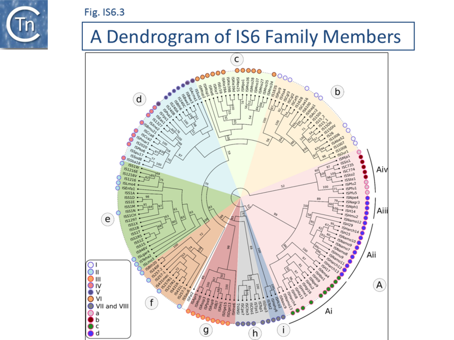
A phylogenetic tree based on the transposase amino acid sequence of the ISfinder collection (Fig. IS6.3) shows that the IS6 family members fall into a number of well-defined clades. This slightly more extensive set of IS corresponds well to the results of another wide-ranging phylogenetic analysis [31]. These clades include one which groups all archaeal IS6 family members (Fig. IS6.3 a) composed mainly of Euryarchaeota (Halobacteria ; Fig. IS6.3 ai-iii). Group aiv includes both Euryarchaeota (Thermococcales and Methanococcales) and Crenarchaeota (Sulfolobales). Of the 10 clades containing bacterial IS: clade b includes examples from the Alpha-, Beta-, and Gamma-proteobacteria, Firmicutes, Cyanobacteria, Acidobacteria and Bacteroidetes ; clade c is more homogenous and is composed of Alphaproteobacteria (Rhizobiaceae and Methylobacteriaceae); clade d includes some Actinobacteria, Alpha-, Beta-, and Gamma-proteobacteria ; while clades e, f, g and h are composed exclusively of Firmicutes (almost exclusively Lactococci in the case of clades e and f). Clades I and j are more mixed.
Clearly, the ISfinder collection does not necessarily reflect the true IS6 family distribution and these grouping should be interpreted with care. For example, although many do not form part of the ISfinderdatabase, IS6 family elements are abundant in archaea and cover almost all of the traditionally recognized archaeal lineages (methanogens, halophiles, thermoacidophiles, and hyperthermophiles [32] (Fig. IS6.3) .
Terminal Inverted Repeats.
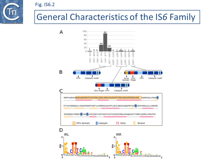
The division into clades is also underlined to some extent by the IR sequences. As shown in Fig. IS6.2 (bottom), in spite of the wide range of bacterial and archaeal species in which family members are found, there is a surprising sequence conservation. In particular, the presence of a G dinucleotide at the IS tips and cTGTt and caaa internal motifs. Sequence motifs are more pronounced when each clade is considered separately (Fig. IS6.4).
- Clade b
(n=16; Actinobacteria, Alpha-, Beta-, and Gamma-proteobacteria) includes a well conserved GG..cTGTTGCAAA signature with little conservation further into each end.
- Clade c
(n= 14; Alphaproteobacteria: Rhizobiaceae and Methylobacteriaceae) shows considerable conservation of an extended motif (GGG... TGTCGCAAA) and some conservation further into both IRL and IRR, although these are different for each end.
- Clade d
(n=24; with Alpha-, Beta-, and Gamma-proteobacteria, Firmicutes, Cyanobacteria, Acidobacteria and Bacteroidetes) maintains stronger traces of parts of these motifs (GG.. tcTGtt and CAaa).
- Clade e
(n=23; s composed mainly of IS from Lactococcus, a single Leuconostoc and other bacilli (Lysteria, Enterococcus);
- Clade f
(n = 11; largely Staphylococci with 2 B. thuringiensis) also exhibit the typical GGTTCTGTTGCAAAGTTt signature and some internal conservation in IRL.
- Clade g
(n = 10; is more heterogenous (Alpha proteobacteria: Methylobacterium, Paracoccus, Roseovarius, Rhizobium, Bradyrhizobium ; Deinococci and Halobacteria). It contains a poorly conserved IR sequence but does include a prominent gG dinucleotide tip and a poorly pronounced tgtcaagtt signature).
- Clade h
(n= 5; composed entirely of Firmicutes (Natranaerobius, Clostridium and Thermoanaerobacter) exhibits a moderately well-defined internal signature TcTgTtAAgTt).
- Clade i
Finally, clade I (n=3) is composed of Halanaerobia and Thermoanaerobacter.
The archaeal-specific clades also generally exhibit well-defined consensus sequences.
- Clade Ai
Is composed of diverse Halobacterial species (Halohasta, Haloferax, Natrinema, Natrialba, Halogeometricum, Natronomonas, Natronococcus, and Haloarcula): GgcACtGTCTAGtT.
- Clade Aii
(n = 12) is composed uniquely of Halobacterial Euryarchaeota with a ggtaGTGTTcagatAaG signature and significant internal conservation which is different for each end.
- Clade Aiii
(n = 5), is composed entirely of Halobacterial Euryarchaeota (Haloarcula, Halomicrobium, Natronomonas, Natronobacterium, Natrinema) also has well conserved ends, ggtcgTGTTTaGTT, and significant internal conservation which is different for each end.
- Clade Aiv
(n = 9) which includes both Euryarchaeota and Crenarchaeota, has poor conservation although on further analysis, an alignment shows significant conservation in the Sulfolobus and in the Pyrococcus groups with good interior conservation also in the 3 Pyrococcal members. It is possible that the IS ends in the Sulfolobus members have not been accurately identified.
MCL analysis [33] for the entire group of transposases using the criteria of ISfinder for classification (IS identification) [34] showed that all members fell within the definition of a single family (Inflation factor 1.2, score >30) and fell into 3 groups: clades b-I; clades Ai-Aiii; and Aiv using the appropriate filter (Inflation factor 2, score >140). The answer to the recent question “An analysis of the IS6/IS26 family of insertion sequences: is it a single family?”[31] is therefore “Probably, yes” according to the ISfinder definition.
A recent study [35] identified a number of IS26 variants with specific mutations in their Tpases. In particular one variant, originally called IS15Δ/IS15D [5][36] was observed to exhibit enhanced activity and it was suggested that such mutants, even though they satisfy ISfinder criteria attributing a new name for an IS (< 95% nucleotide identity and/or < 98% amino acid identity). It has been suggested that such variant should be suffixed as IS26.v1, .v2 etc. [35]. This makes sense if the mutation is not functionally neutral results in a change IS properties or behavior.
Genomic Impact and Clinical Importance
Activity resulting in horizontal dissemination is suggested, for example, by the observation that copies identical to Mycobacterium fortuitum IS6100 [37] (Clade b) occur in other bacteria: as part of a plasmid-associated catabolic transposon carrying genes for nylon degradation in Arthrobacter sp. [38], from the Pseudomonas aeruginosa plasmid R1003 [39], in integrons of the In4-type integrons from transposons such as Tn1696 [40][41] and within the Xanthomonas campestris transposon Tn5393b [42]. Similar copies have also been reported in Salmonella enterica (typhimurium) [43], and on plasmid pACM1 from Klebsiella oxytoca (AF107205) [44].
Passenger Genes
A number of IS families contain members, called tIS which carry passenger genes. A single member of the family, ISDsp3, present in a single copy in Dehalococcoides sp. BAV1 carries a passenger gene annotated as a hypothetical protein..
Expression of neighboring genes
The formation of hybrid promoters on insertion, where the inserted element provides a -35 promoter component and the flanking sequence carries a -10 promoter component, is clearly a general property of members of the IS6 family [22][45][46][47][48][49].
For example, IS257 [50](Clade f) (also known as IS431) has played an important role in sequestering a variety of antibiotic resistance genes in clinical isolates of methicillin resistant Staphylococcus aureus (MRSA) (e.g. [45][46][51][52]. It provides an outward oriented promoter which drives expression of genes located proximal to the left end. Moreover, both left and right ends appear to carry a –35 promoter component which would permit formation of hybrid promoters on insertion next to a resident –10 element [46][53]. Insertion of can result in activation of a neighboring gene using both a hybrid promoter and an indigenous promoter [46]. IS257 is also involved in expression of tetA [49] and dfrA [45] in S. aureus.. This is also true of IS26 which forms hybrid promoters shown to drive antibiotic resistance genes such as aphA7 ( Pasteurella piscicida [54] Klebsiella pneumoniae [22]), blaSHV-2a (Pseudomonas aeruginosa [55]) and wide spectrum beta-lactam resistance gene blaKPC [56][57]. While IS6100 [37] (Clade b) drives strA strB expression in X. campestris pv. vesicatoria [42]
The formation of hybrid promoters on insertion (Table IS and Gene Expression) is clearly a general property of members of the IS6 family [22][45][46][47][48][49].
Pseudo-compound transposons
This IS family is able to form transposons which resemble compound transposons with the flanking IS in direct repeat but, because of the particular transposition mechanism of IS6 family members which involves the formation of cointegrates (see below), were called pseudo-compound transposons [2][16]. These include Tn610 (flanked by IS6100 [37]), Tn4003 and others (flanked by IS257 [51][58][59]), Tn2680[7] (flanked by IS26 [60]).
IS26 and the Clinical Landscape
In view of the particular importance of IS26 in clinical settings it is worthwhile devoting a separate section to the contribution of this IS to the clinical landscape. IS26 [7][8] (clade b) is encountered with increasing frequency in plasmids of clinical importance where it is involved in: sequestering antibiotic resistance genes and generating arrays of these genes in clinically important conjugative plasmids and in the host chromosome; expression of antibiotic resistance genes; and other plasmid rearrangements (see [28][59][61][62][63][64][65][66]).
Recognition of its place as an important player has derived from the large number of sequences now available of multiple antibiotic resistance plasmids and chromosomal segments such as Genomic Resistance Islands (GRI). It is now no longer practical to provide a complete analysis of the literature (A PubMed search (19th November 2020) using IS26 as the search term yielded nearly 450 citations). The references in the following are not exhaustive but simply provide examples.
IS Arrays
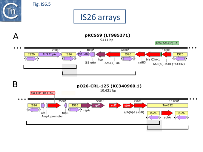
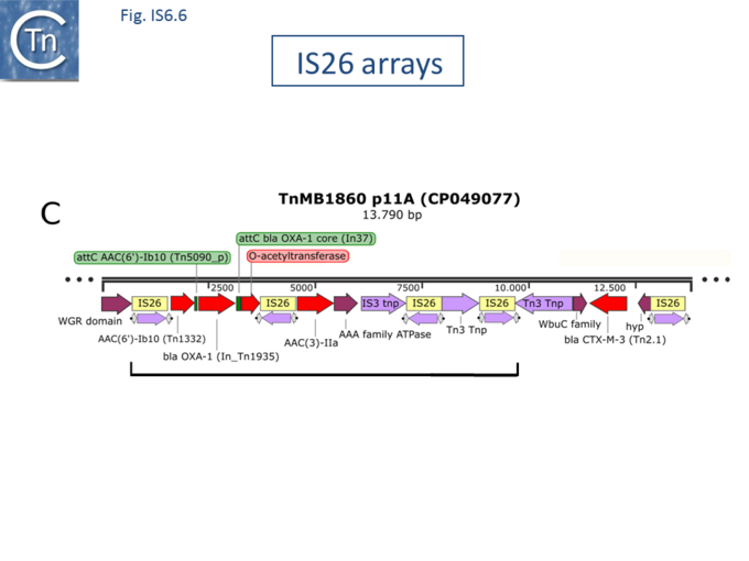
IS6 family members are often found in arrays (Fig. IS6.5 and Fig. IS6.6) in direct and inverted repeat in multiple drug resistant plasmids (e.g. Salmonella. typhimurium [28][60][67], Klebsiella quasipneumoniae [68], Acinetobacter baumannii [64][69], Proteus mirabilis [70] and uncultured sewage bacteria [71] (among many others). These are often intercalated in or next to other transposable elements rather than neatly flanking Antibiotic Resistance genes and can form units able to undergo tandem amplification.
IS26-mediated Gene Amplification
Early studies with Tn1525 (from Salmonella enterica serovar Panama), in which an aphA1 (aph (3') (5")-I) gene is flanked by two directly repeated copies of a the IS6 family member, IS15, reported tandem amplification of aphA1 when the host was challenged by kanamycin [74]. Restriction enzyme mapping was used to demonstrate that the amplified segments were of the type IS-aph-IS-aph-IS-aph-IS but no direct sequence data is available. Amplification was thought to occur by homologous recombination between two flanking IS15 copies since it occurred in a wildtype host but the transposon was stable in a recA genetic background. Another example was observed following treatment of a patient with Tobramycin in clinical isolates of Acinetobacter baumannii from a single patient over a period of days with continued antibiotic treatment [75]. Amplification occurred with Tn6020, an IS26-based transposon in which the flanking IS bracket a similar aphA1 gene and could also be reproduced in bacterial culture [75]. In this case, the amplified unit was proposed to be IS-aph-IS-IS-aph-IS-IS-aph-IS. This structure would clearly be unusual but may be due to a misinterpretation of the depth of coverage of the region. In addition, the amplified transposon had inserted into a known target prior to amplification generating the expected eight base pair target repeat but an 8bp segment between the first DR and the first IS end (DR-8 bp-ISaph-IS-IS-aph-IS-ISaph-IS…DR). A third example [73] was identified during a study of clinical isolates of non-carbapenemase-producing Carbapenem-Resistant Enterobacteria, non-CP-CRE, isolated from several patients with recurrent bacteraemia. An increase in carbapenem resistance occurred partially due to IS26-mediated amplification up to 10 fold of a DNA segment carrying blaOXA-1 and blaCTX-M-1 genes These form part of a larger chromosomal structure of IS26 arrays which they call TnMB1860 (Fig. IS6.6). It was unclear whether this cassette amplification was due to transposition activity or, as had been observed in similar, IS1-mediated, gene amplifications [76][77][78][79][80][81] which may occur by replication slippage between direct repeats or by unequal crossing-over [82][83].
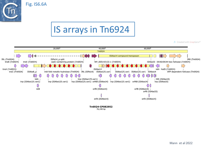
A more dramatic amplicon, was observed in the case of the IS6 family member ISAba125 in the transposon Tn6924 (Fig.IS6.6A) in which two blocks of 7 and 8 IS arrays are present flanking 14 copies of aminoglycoside resistance APH(3')-VI genes (aka aphA6, amikacin resistance) which gives rise to high levels of resistance, presumably facilitated by high amikacin selective pressure [84].
Another example has been revealed by Hubbard et al [85] who analysed a multi resistant derivative of the clinically important, globally dispersed pathogenic, Escherichia coli ST131 subclade H30Rx, isolated from a number of bacteraemic patients and revealed that increased piperacillin/tazobactam resistance was due to IS26-mediated amplification of blaTEM-1B. A similar type of limited (tandem dimer) amplification of an IS26-flanked blaSHV-5-carrying DNA segment found in plasmids from a number of geographically diverse enteric species was identified in a nosocomial Enterobacter cloacae strain [86]. More extensive amplification (>10 fold) was observed with the same DNA segment located in a different plasmid in a well-characterised laboratory strain of Escherichia coli and occurred in a recA-independent manner [62] while even higher levels of tandem amplification (~65 fold) of the aphA1 gene in the IS26-based Tn6020 were identified in Acinetobacter baumannii [75].
Extensive IS26-mediated drug resistance gene amplification has also recently been observed experimentally under controlled laboratory conditions during growth of the host strain in sub-lethal concentrations of meropenem or tobramycin and gave rise to high carbapenem resistance [87].
IS26-mediated Plasmid Cointegration
The earliest studies on this family of IS demonstrated that they could generate cointegrates as part of the transposition mechanism (see Cointegrate formation below) [6][7][9][12][13][30].
Several studies have now demonstrated that this can occur in a clinical setting. For example, plasmid pBK32533 (KP345882) [88], carried by E. coli BK32533 isolated from a patient with a urinary tract infection is an IS26-mediated cointegrate between Klebsiella pneumoniae BK30661 plasmid pBK30661 (KF954759) [89] and a relative of Salmonella enterica p1643_10 (KF056330) [90]. Interestingly, the flanks of the IS26 copies at the junction of the two plasmids are TGTTTTTT-IS-TTATTAAT and TTATTAAT-IS-TGTTTTTT. The most parsimonious explanation would be that pBK32533 was generated in a multi-step inter-molecular transposition event: in one step, an IS26 copy from an unknown source used a TTATTAAT target sequence in pBK30661 and this cointegrate was then resolved resulting in pBK30661 containing an IS26 copy flanked by the target repeat (TTATTAAT-IS26-TTATTAAT) and, in a second step, a TGTTTTTT sequence in p1643_10 was targeted by the pBK30661 IS26 to generate the final cointegrate in which the two IS26 copies are flanked by the observed target sequences. Additional examples have been identified in KPC-producing Proteus mirabilis [70] and in Klebsiella pneumoniae also involving inversions [66][91].
Organization
IS6 family members range in length from 696 to 1761 bp (ISFinder) with the majority between 789 bp (IS257) to 880 bp (IS6100) (Fig. IS6.2 A) and generally create 8 bp direct flanking target repeats (DR) on insertion [7].
The transposase
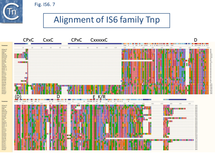
A single transposase orf is transcribed from a promoter at the left end and stretches across almost the entire IS. The putative transposases (Tpases) are between 234 (IS15Δ) and 254 (e.g., IS6100) amino acids long with a majority in the 220-250 amino acid range. They are very closely related and show identity levels ranging from 40 to 94% with a helix-turn-helix (HTH) and a typical catalytic motif (DDE) (Fig. IS6.2 C and Fig. IS6.7). However, the 7 members of clade h, all from Clostridia, are somewhat larger than other IS6 family members (approximately 1200bp, Fig. IS6.2 A) with longer transposases (340-350 amino acids) as a consequence of an N-terminal extension with a predicted Zinc Finger (ZF) composed of several CxxC motifs (Fig. IS6.2 B; Fig. IS6.7). A Blast analysis of the non-redundant protein database at NCBI revealed a large number of IS6 family transposases of this type (data not shown). The vast majority of these were from Clostridial species. In addition, the transposases of members of clade i (450 amino acids) have both the ZF domain and a supplementary N-terminal extension.
Several members (e.g. ISRle39a, ISRle39b and ISEnfa1) apparently require a frameshift for Tpase expression. It is at present unclear whether this is biologically relevant. However, alignment with similar sequences in the public databases suggests that ISEnfa1 itself has an insertion of 10 nucleotides and is therefore unlikely to be active.
Transposase expression
In the case of IS26, the promoter is located within the first 82 bp of the left end and the intact orf is required for transposition activity [8], and the predicted amino acid sequence of the corresponding protein exhibits a strong DDE motif (Fig. IS6.2 C; Fig. IS6.7) Translation products of this frame have been demonstrated for IS240 [26]. Little is known concerning the control of transposase expression although transposition activity of IS6100 in Streptomyces lividans [92] is significantly increased when the element is placed downstream from a strong promoter. This is surprising since IS generally incorporate mechanisms to restrict transposition induced by insertion into highly transcribed genes (see Fig 1.32.1).
Terminal Inverted Repeats
All carry short, related terminal IR of 13- 25bp. (depending on how mismatches are scored ). As shown in Fig. IS6.2 D, in spite of the wide range of bacterial and archaeal species in which family members are found, there is a surprising sequence conservation. In particular, the presence of a G dinucleotide at the IS tips and cTGTt and caaa internal motifs (where uppercase letters are fully conserved and lowercase letters are strongly conserved nucleotides). Sequence motifs are more pronounced when each clade is considered separately (Fig. IS6.4).
Mechanism: the state of play
Early studies suggested that IS6 family members give rise exclusively to replicon fusions (cointegrates) in which the donor and target replicons are separated by two directly repeated IS copies
(e.g. IS15Δ/IS15D [6], IS26/IS46/IS140 [7][9][11], IS257[53], IS1936 [13], ISS1 [93]). More recent results principally with IS26 have suggested that this IS perhaps like IS1 (IS1 family) [94] and IS903 (IS5 family) [95][96], members of this family may be able to transpose using alternative pathways [16][97][98][99].
Cointegrate formation
Transposition of IS6 family elements to generate cointegrates [6][9][11][12] presumably occurs in a replicative manner by Target Primed Transposon Replication (For a discussion see “Influence of transposition mechanisms on genome impact”; Fig 17.1 and Fig. 17.2), and as expected, requires two functional IS26 ends [100]. As shown in Fig. IS6.8 (top), intermolecular replicative transposition of this type generates fused donor and target replicons which are separated by two copies of the IS in direct repeat at the replicon boundaries. The initial Direct Repeats (DR) flanking the donor IS are distributed between each daughter IS in the cointegrate as is the DR generated in the target site. Recombination between the two IS then regenerates the donor molecule with the original DRs and a target molecule in which the IS is flanked by new DR. No known specific resolvase system such as that found in Tn3-related elements (see IS Derivatives of Tn3 family transposons) has been identified in this family but “Resolution” of IS6-mediated cointegrates was observed to depend on a functional recA gene in several cases and therefore occurs using the host homologous recombination pathway [6][9].
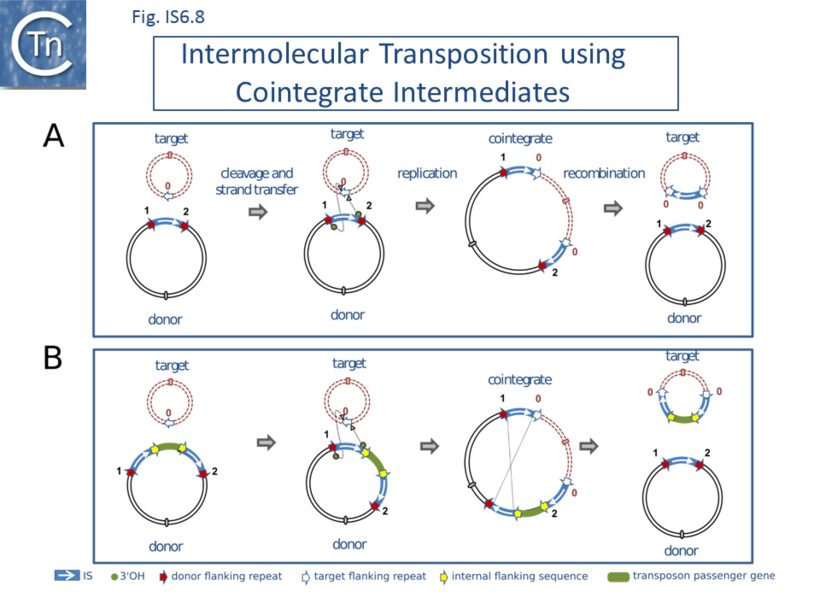
A systematic analysis of the cointegrate forming properties of an artificial IS26-based pseudo-compound transposon with a chloramphenicol transacetylase passenger gene has demonstrated that if the inside ends of the two flanking IS are ablated, the full-length transposon can promote cointegrate formation at a low frequency. The sequence of the resulting cointegrates confirmed that the donor and target replicons were separated by a copy of the entire transposon at each junction with the appropriate 8 base pair target duplication (He et al., pers. comm).
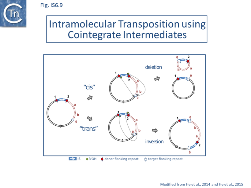
While the intermolecular cointegrate pathway leads to replicon fusion, transposition can also occur within the same replicon. Intramolecular transposition using the replicative mechanism gives rise to deletion or inversion of DNA located between the IS and its target site. The outcome depends on the orientation of the two attacking IS ends (Fig. IS6.9). Intramolecular transposition of this type can explain the assembly of antibiotic resistance gene clusters (e.g. [66]).
IS6 family members are known to generate structures that resemble composite transposons in which a passenger gene (such as a gene specifying antibiotic resistance) is flanked by two IS copies. Generally, other flanking IS in compound structures can occur as direct or inverted repeat copies (IS history; Fig 2.3, Fig. 2.4). However, in the case of IS6 functional “compound transposons”, the flanking IS are always found as direct repeats. This is a direct consequence of the (homologous) recombination event required to resolve the cointegrate structure [6][9]. As shown in Fig. IS6.8 (bottom) [102], transposition is initiated by one of the flanking IS to generate a cointegrate structure with three IS copies. “Resolution” resulting in transfer of the transposon passenger gene requires recombination between the “new” IS copy and the copy which was not involved in generating the cointegrate. The implications of this model [2][102] are that the transposon passenger gene(s) are simply transferred from donor to target molecules in the “resolution” event and are therefore lost from the donor “transposon”. Clearly this pathway could initiate from a donor in which the flanking IS6 family members were inverted with respect to each other. However, transposition would be arrested at the cointegrate stage because there is no suitable second IS to participate in recombination. It is for this reason that compound IS6-based transposons carry directly repeated flanking IS copies. These previously published models (e.g.[2][66][91][102] have been revisited and it has been recently proposed [16] that the term pseudo-compound transposons first used over 30 years ago [2] should be resurrected to describe these IS6 family structures.
Circular transposon molecules: translocatable units (TU)
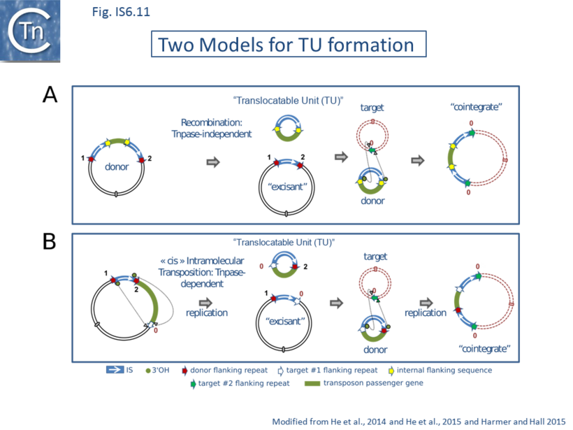
Although IS26 transposition appears to be replicative with formation of cointegrate molecules, results from in vivo experiments suggest that its transposition may be more complex [99]. The idea that IS26 might mobilize DNA in an unusual way arose from the observation that IS6 family members can often be found in the form of arrays [98][99] which could be interpreted as overlapping pseudo-compound transposons [16] (Fig. IS6.5). Note that IS26 and potential IS26-based transposons do not necessarily carry flanking direct target repeats but, as is the case for other TE, which transpose by replicative transposition such as members of the Tn3 family, intramolecular transposition would lead to loss of the flanking repeats (Fig. IS6.9). This led to the suggestion that IS26 might be able to transpose via a novel circular form called Translocatable Units (TU) [98][99] (not to be confused with those originally described in the sea urchin and other eukaryotes [103]) such as those shown in Fig. IS6.10. These potential circular transposition intermediates which were proposed to include a single IS26 copy along with neighboring DNA are structurally similar to IS1-based circles observed in the 1970s (e.g. [76][79]). Translocatable units differ from the transposon circles identified during copy-out-paste-in transposition by IS of the IS3 (Fig. IS3. 9A; IS3 family transposition pathway), IS21 (Fig. IS21.7), IS30, IS256 and ISL3 families where the circular IS transposition intermediate has abutted left and right ends separated by a few base pairs and is extremely reactive to the cognate transposase. In stark contrast, for IS26, the IS ends would be separated by the neighboring DNA sequence rather than by a few base pairs (Fig. IS6.11).
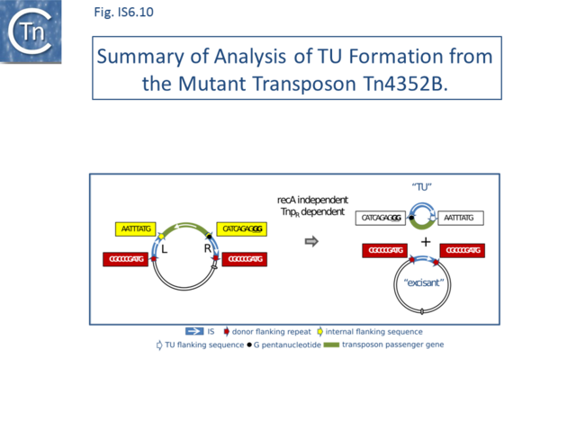
Evidence for the excision step of translocatable units was obtained [98] from the study of the stability of two IS26-based pseudo-compound transposons, “wildtype” Tn4352 [25] and “mutant” Tn4352B [104] which carry the aphA1 gene specifying resistance to kanamycin. Tn4352B is a special mutant derivative of Tn4352 including an additional GG dinucleotide at the left internal end of one of the component IS26 copies to generate a string of 5 G nucleotides at the IS tip which appears to render the transposon unstable. Cells carrying the plasmid lose the resistance gene from the mutant Tn4352B at an appreciable rate in the absence of selection. This generates a “donor” plasmid with one copy of IS26 flanked by the original Tn4352B-associated 8bp direct repeats and an excision product with the size expected for a TU containing the second IS flanked by the sequences of the original central segment presumably including the additional GG dinucleotide together with the aphA1 gene. TU formation, as judged by a PCR reaction, appeared to be dependent on the GG insertion (since, surprisingly, no TU could be detected from the wildtype Tn4352) but not on the surrounding sequence environment. Excision required an active transposase. In a test in which the target plasmid also carried an IS26 copy (a targeted integration reaction – see below), there appeared to be no difference in cointegrate formation frequencies between single IS26 copies with or without the additional GG dinucleotide. However, results from a standard integration test into a plasmid without a resident IS26 copy were not reported. The excision process occurs in a recA background and therefore does not require the host homologous recombination system. Moreover, frameshift mutations in both IS, which should produce severely truncated transposase, eliminated activity. This implies that the process is dependent on transposition. However, excision continued to occur if the transposase of the GG-IS copy was inactivated but was eliminated when the same transposase mutation was introduced into the ”wildtype” IS copy. This is curious since it implies that the IS26 transposase must act exclusively in cis on the IS from which it is expressed (see Co-translational binding and multimerization).
A summary of these results is shown in Fig. IS6.10. These data suggest that excision is driven by the wildtype IS26 (L), leaving the right hand IS in the excisant. At present, there is no obvious mechanistic explanation for this phenomenon. It should be noted that recombination between directly repeated copies of IS1 which flank the majority of Antibiotic Resistance genes in the plasmid R100.1 (NR1) generates a non-replicative circular molecule, the r-determinant (r-det), with a single IS1 copy. In this case too, this “constitutive” circle production is due to a (uncharacterized) mutation in the plasmid, although in this case, circle production requires recA [105].
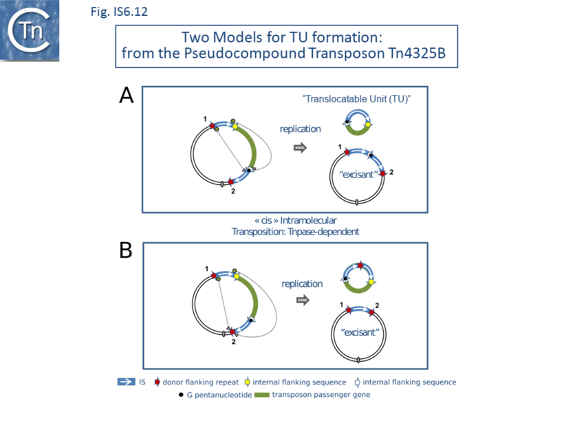
However, “Classical” recombination and transposition models do not fit the data The results appear to rule out two obvious models (Fig. IS6.11): since, although both would generate the correct TU and “excisant”, the first (top panel) requires homologous recombination between two directly repeated IS26 copies (mechanistically equivalent to the “resolution” step in intermolecular IS6 transposition) and the second (bottom panel), which requires a functional transposase as observed [98][99], would not generate the correct flanking sequences. Modification of the transposition model to take into account the entire transposon (Fig. IS6.12) in which the active IS26L uses either of flanking sequences of IS26R does not generate the correct structures. Thus the observed structures must be generated by another, and at present unknown, pathway. One possibility is that TU are generated by reversing a non-replicative targeted insertion mechanism presented below (Fig. IS6.14; see Targeted Transposition).
Tn4352B Instability is Dependent on Specific-recA Alleles.
Curiously, although a Tn4352B copy carried by a low copy number IncC plasmid was stable in two different K. pneumoniae hosts [106] and in two recombination proficient E. coli hosts [107], it became unstable in certain recA-deficient E. coli hosts. The instability appeared to depend on the particular recA allele involved: it occurred in the presence of the recA1 allele [108] but not in the presence of recA13 [109] or of a novel recA mutant fortuitously identified during the analysis [107].
The necessity of a specific defective RecA protein derivative to accomplish the formation of a proposed TU transposition intermediate is intriguing and certainly merits further investigation.
To summarize: It has been clearly demonstrated that circular DNA species carrying a single IS26 copy together with flanking “passenger” DNA can be generated efficiently in vivo from a variant plasmid replicon [104] and also that replicons carrying a single IS26 copy are capable of integrating into a second replicon to form a cointegrate. This occurs at a frequency 102-fold higher if the target plasmid contains a single IS copy and in a targeted manner not involving IS duplication.
The TU insertion pathway was addressed by transforming TU, constructed in vitro taking advantage of a unique IS26 restriction site, into recombination deficient cells carrying an appropriate target plasmid [97]. Establishment of the aphA1-carrying TU was dependent on the presence of a resident plasmid carrying an IS26 copy and occurred next to the resident IS26 copy. The DNA of two TU each with a different antibiotic resistance gene was shown to undergo this type of targeted integration and, moreover, were able to consecutively insert to generate a typical IS26 array. It was also shown that the TU circles produced from the mutant Tn4352B were able to subsequently undergo insertion [96]. Therefore, artificially produced TU and those produced from Tn4352B are capable of subsequent insertion. In addition, Zienkiewicz et [66] have presented evidence that “an IS26 -bla SHV-5 module that was capable of amplification in cis could also move in trans and concluded that: “consistent with earlier observations [18][61][110], it did not copy by transposition but via IS26-mediated recombination.
Targeted transposition.
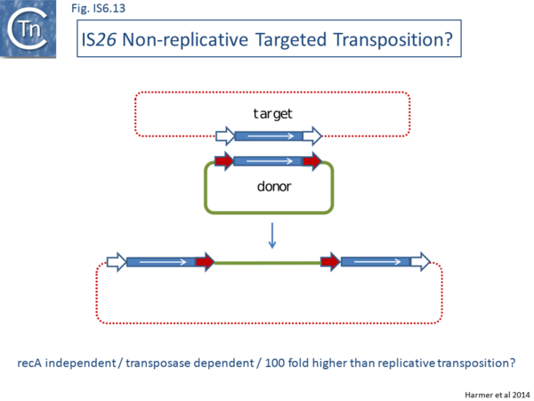
Targeted IS26 transposition, was also observed in intermolecular cointegrate formation where cointegrate formation frequency was significantly increased about 100 fold if the target replicon also contained an IS26 copy [99]. A similar result was obtained in Escherichia coli with a related IS, IS1216 [111] whereas a third member of the family, IS257 (e.g., IS431) showed a much lower level of activity using the same assay. As for TU integration, this phenomenon does not appear to be the result of homologous recombination between the IS copies carried by donor and target molecules since the reaction was independent of RecA. Using a PCR-based assay to identify the replicon fusions between IS26-containing donor and target plasmids, it was observed that the resulting cointegrate (Fig. IS6.13) did not contain an additional copy of IS26 which would be expected if replicative transposition were involved (Fig. IS6.12). This suggests that the phenomenon results from a conservative recombination mechanism. Despite the absence of RecA, the observed cointegrate is structurally equivalent to the recombination product between the two IS26 copies in the donor and target plasmids. However, it indeed appears to be transposition related since the phenomenon requires an active transposase in both donor and target replicons [99]. When each of the triad of conserved DDE residues were mutated individually in the donor plasmid, the targeted insertion frequency decreased significantly.
Another characteristic of the products was that the flanking 8 bp repeats carried by the donor and recipient IS26 copies are in some way exchanged [99] (Fig. IS6.13). This suggests a model in which transposase might catalyze an exchange of flanking DNA during the fusion process.
A Model for Targeted Integration
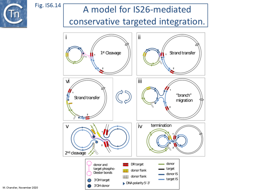
One possibility (Fig. IS6.13) is that two IS ends from different IS copies in separate replicons are synapsed intermolecularly in the same transpososome (Fig. IS6.14 i). Strand exchange would then couple the donor and target replicons (Fig. IS6.14 ii). A similar mechanism has been invoked to explain “targeted” insertion of IS3 and IS30 family members into related IRs (Fig. IS3.14) [112][113]. Branch migration (Fig. IS6.14 iii) would lead to exchange of an entire IS strand (Fig. IS6.14 iv) and cleavage at the distal IS end and strand transfer (Fig. IS6.14 v) would result in the observed cointegrate (Fig. IS6.14 vi) containing a single strand nick on opposite strands at each end of the donor DNA molecule. These could simply be repaired or eliminated by plasmid replication. Each IS would be composed of complementary DNA strands from each of the original donor and target IS copies. This proposed mechanism would retain the DNA flanks of the IS in the original target replicon, be dependent on an active transposase and independent of the host recA system. It seems probable that mismatches between the two participant IS would inhibit the strand migration reaction. This may be the reason for the observation that introducing a frameshift mutation by insertion of additional bases into the transposase gene of either participating IS26 copy reduces the frequency of targeted cointegration [99] since, not only does this produce a truncated transposase but also introduces a mismatch. As in the case of intermolecular targeting of the IS3 family member, IS911 [114], might require the RegG helicase to promote strand migration.
The model shown in Fig. IS6.14 presents the transposition process as a progression involving two consecutive, temporally separated, strand cleavages interrupted by a strand migration. However, it seems equally probable that both cleavage reactions are coordinated within a single transpososome (The Transpososome) including both donor IS ends and the target IS ends. This would be compatible with the known properties of trans cleavage of several transposases in which a transposase molecule bound to one transposon end catalyses cleavage of the opposite end (Cleavage in Trans: A Committed Complex). Recently, evidence have been presented supporting this type of “rolling in” model [100]. In addition, it has been observed that targeted integration does not necessarily require two functional ends of the donor IS [115]. This indicates the strand migration process could be terminated (probably via HJ resolution) prior to encountering the second IR. Moreover, using two IS, IS1006 and IS1008 [116] which have significant identity to IS26, at their ends together with a hybrid molecule IS1006/1008 constructed in vitro, it was shown that targeted integration required both identical transposases and identical DNA sequences at the reacting ends. The authors propose a model in which a single IS end is cleaved and transferred to the flank of the target IS end, an event which creates a Holliday junction (HJ) which, on branch migration, is resolved. This differs from the model shown here (Fig. IS6.14) since it does not involve transposase-mediated cleavage at the second IS end. It is similar to that proposed for targeted insertion of IS911 [112][114][117][118] which requires the RecG helicase and, presumably RuvC. However, it has now been demonstrated that neither recG, ruvA or ruvC genes influence this type of IS26 activity. This therefore implies that HJ structures, if they are indeed formed, must be processed by a different pathway, as yet to be deciphered [119].
Emerging Biochemistry: Transposase binding.
Some initial studies have begun to address binding of the IS26 transposase, Tnp26, to its cognate IS [120]. Tnp26 carries a typical DDE motif with the conserved triad located at codons 82, 142 and 177 (Fig. IS6.7) and, in addition, includes the often conserved K/R residue 7 amino acids downstream from the final E(177) (Major Groups are Defined by the Type of Transposase They Use). As expected, the DDE triad is required for transposition activity [99].
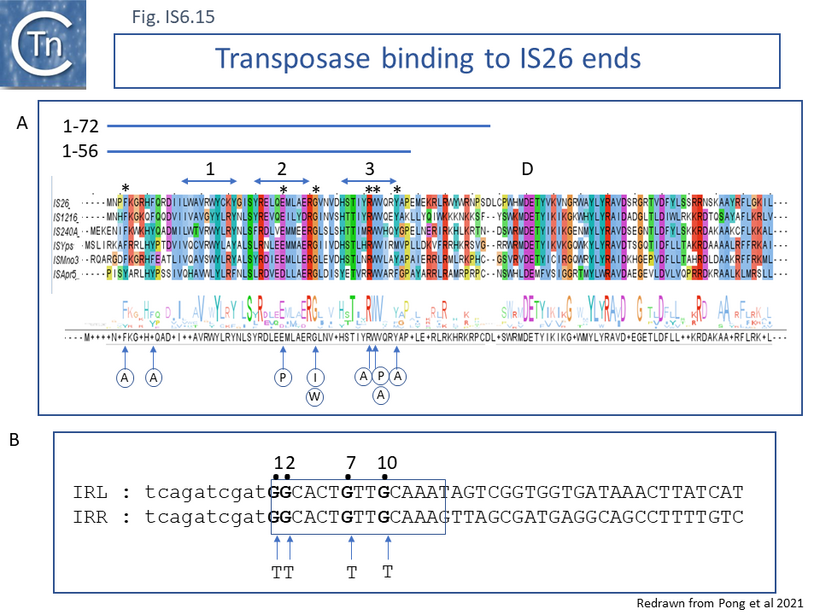
Moreover, the DNA binding domain of a number of transposases of different IS families is located in the N-terminal region which often contains multiple alpha-helices, in particular, helix-turn-helix motifs (e.g. IS1 family, IS3 family, IS30 family, IS1595 family, IS256 family). This analysis [120] identified a potential N-terminal triple helix (Fig. IS6.15 A).
The binding studies used the full length 234 amino acid (27.9kD) Tnp26 or various fragments cloned as a fusion with Maltose binding protein (MBP) and were used with the MBP tag since its removal resulted in precipitation. The Tnp fragments composed of amino acids 1-56 (Tnp1-56, 6.95kD) and 1-72 (Tnp1-72, 9.11kD) were chosen following a secondary structure prediction of the transposase. They contain the potential triple helix. This results in fusion proteins of 72.6kd, 51.6 kD and 53.7 kD respectively.
TheMBP-tagged proteins were used in EMSA assays with 50bp Cy5-labeled double strand DNA fragments containing the 14 bp IRs (Fig. IS6.15 B). The wildtype tnp-MPB and tnp1-56 fusions generated a relatively weak retarded single band with both IRL and IRR, consistent with binding to the IR. That obtained with tnp1-56 was significantly weaker than the wildtype while Tnp1-72 failed to exhibit the complex. At higher protein concentrations, DNA was retained in the well a phenomenon which did not occur with “random” DNA and could represent paired end complexes or simply sequence-specific aggregation. A number of mutations in Tnp26 including two within helix 3 and two within a few residues of the N-terminal end (Fig. IS6.15 A) eliminated the retarded complex as well as reducing the material in the well.
Each of the Tnp mutations were observed to reduced cointegrate formation in vivo by over 2 orders of magnitude in vivo.
To determine the IR nucleotides important for Tnp recognition, a number of mutations were introduced in the IRs (Fig. IS6.15 B). A double mutant (GG>TT) at the IR tip only slightly affected complex formation as might be expected since in general, the ends of several IS have been found to be divided into two functional domains: the tip at which transposition chemistry occurs and a region inside the IR to which transposase binds in a sequence-specific way (IS Organization). A G>T change at position 7 appeared to abolish complex formation whereas a G>T change at position 10 did not.
Clearly, additional and more systematic mutagenesis experiments would be able to define the transposase binding site in more detail and determine whether external DNA might be required to assist in binding in a non-sequence-specific manner. Moreover, it cannot at present be ruled out that the relatively large MBP addition affects overall behavior of transposase and its derivatives. It will be of interest to determine whether Tnp26 can form dimeric or multimeric species and can form one or a number of different paired end complexes (PEC; Transposase expression and activity) with IS26 IR in which two ends are bridged by transposase, whether the wildtype transposase acts as a dimer and how end cleavages occur.
Conclusions and Future Directions
We have presented a survey of our present knowledge concerning the properties, distribution and activities of IS6 family members and their importance, in particular that of IS26, in gene acquisition and gene flow of antibacterial resistance in enterobacteria. There are many questions which remain to be answered and we feel that some care should be exercised in interpreting some of the very interesting results in the absence of formal proof. For example, the notion that the basic IS6 family transposition unit is a non-replicative circular DNA molecule carrying a single IS copy is attractive and would provide a nice parallel to the integron antibiotic resistance gene cassette intermediates [121][122][123] but such a molecule, a TU, has thus far been formally observed in only a single case. It was generated in vivo from an IS26-flanked peudo-transposon in which one of the two flanking IS involved included a mutation and rendered the transposon unstable. The “wildtype” transposon was stable [98]. Since “TU” is now being used in the literature to describe IS26-flanked DNA segments in multimeric arrays (e.g. [85], it is essential to provide more formal evidence that these non-replicative DNA circles are indeed general intermediates in the IS26 transposition pathway and are not simply amplified units (AU). The fact that a replicating plasmid containing a single IS copy is able to form cointegrates does not à priori support a model for TU transposition and is not necessarily simply a TU that has the capacity to replicate [99] although the observation that artificially constructed TU can undergo targeted insertion when introduced into a suitable cell by transformation [97] supports the TU hypothesis. A second important question to be answered is how targeted integration occurs. We have suggested one model and suggested ways it might be tested (Fig. IS6.14). The answers to many of these fascinating outstanding questions will be provided when the biochemistry of the reactions is known.
Acknowledgements
We would like to thank Susu He (Nanjing University) for stimulating discussions concerning the transposition models and for supplying the IS26 transposase secondary structure predictions in Fig. IS6.2 C.
Fred Dyda and Alison Hickman (NIDDK, NIH, Bethesda, Maryland, USA) for reading this chapter and for suggestions and Sally Partridge (The University of Sydney and Westmead Hospital, Australia) for critical comments.
Bibliography
- ↑ Varani A, He S, Siguier P, Ross K, Chandler M . The IS6 family, a clinically important group of insertion sequences including IS26. - Mob DNA: 2021 Mar 23, 12(1);11 [PubMed:33757578] [DOI]
- ↑ 2.0 2.1 2.2 2.3 2.4 2.5 2.6 Galas DJ, Chandler M. Bacterial Insertion Sequences. In: Berg DE, Howe MM, editors. Mob DNA. Washington, D.C.: American Society for Microbiology; 1989. p. 109–162.
- ↑ Berg DE, Davies J, Allet B, Rochaix JD. Transposition of R factor genes to bacteriophage lambda. ProcNatlAcadSciUSA. 1975;72:3628–3632.
- ↑ 4.0 4.1 Labigne-Roussel A, Courvalin P. IS15, a new insertion sequence widely spread in R plasmids of gram-negative bacteria. MolGenGenet. 1983;189:102–112.
- ↑ 5.0 5.1 5.2 Trieu-Cuot P, Courvalin P. Nucleotide sequence of the transposable element IS15. Gene. 1984;30:113–120.
- ↑ 6.0 6.1 6.2 6.3 6.4 6.5 6.6 6.7 Trieu-Cuot P, Courvalin P . Transposition behavior of IS15 and its progenitor IS15-delta: are cointegrates exclusive end products? - Plasmid: 1985 Jul, 14(1);80-9 [PubMed:2994132] [DOI]
- ↑ 7.0 7.1 7.2 7.3 7.4 7.5 7.6 Mollet B, Iida S, Shepherd J, Arber W . Nucleotide sequence of IS26, a new prokaryotic mobile genetic element. - Nucleic Acids Res: 1983 Sep 24, 11(18);6319-30 [PubMed:6312419] [DOI]
- ↑ 8.0 8.1 8.2 8.3 Mollet B, Iida S, Arber W . Gene organization and target specificity of the prokaryotic mobile genetic element IS26. - Mol Gen Genet: 1985, 201(2);198-203 [PubMed:3003524] [DOI]
- ↑ 9.0 9.1 9.2 9.3 9.4 9.5 9.6 Brown AM, Coupland GM, Willetts NS . Characterization of IS46, an insertion sequence found on two IncN plasmids. - J Bacteriol: 1984 Aug, 159(2);472-81 [PubMed:6086571] [DOI]
- ↑ Nucken EJ, Henschke RB, Schmidt FR. Nucleotide-sequence of insertion element IS15 delta IV from plasmid pBP11. DNA Seq. 1990;1:85–88.
- ↑ 11.0 11.1 11.2 Bräu B, Piepersberg W . Cointegrational transduction and mobilization of gentamicin resistance plasmid pWP14a is mediated by IS140. - Mol Gen Genet: 1983, 189(2);298-303 [PubMed:6304469] [DOI]
- ↑ 12.0 12.1 12.2 Nies BA, Meyer JF, Wiedemann B . Tn2440, a composite tetracycline resistance transposon with direct repeated copies of IS160 at its flanks. - J Gen Microbiol: 1985 Sep, 131(9);2443-7 [PubMed:2999303] [DOI]
- ↑ 13.0 13.1 13.2 13.3 Colonna B, Bernardini M, Micheli G, Maimone F, Nicoletti M, Casalino M. The Salmonella wien virulence plasmid pZM3 carries Tn1935, a multiresistance transposon containing a composite IS1936- kanamycin resistance element. Plasmid. 1988;20:221–231.
- ↑ 14.0 14.1 Barg NL, Register S, Thomson C, Amyes S . Sequence identity with type VIII and association with IS176 of type IIIc dihydrofolate reductase from Shigella sonnei. - Antimicrob Agents Chemother: 1995 Jan, 39(1);112-6 [PubMed:7695291] [DOI]
- ↑ Hall RM . pKM101 is an IS46-promoted deletion of R46. - Nucleic Acids Res: 1987 Jul 10, 15(13);5479 [PubMed:3037494] [DOI]
- ↑ 16.0 16.1 16.2 16.3 16.4 Harmer CJ, Pong CH, Hall RM . Structures bounded by directly-oriented members of the IS26 family are pseudo-compound transposons. - Plasmid: 2020 Sep, 111;102530 [PubMed:32871211] [DOI]
- ↑ Correia FF, Inouye S, Inouye M . A family of small repeated elements with some transposon-like properties in the genome of Neisseria gonorrhoeae. - J Biol Chem: 1988 Sep 5, 263(25);12194-8 [PubMed:2842323]
- ↑ 18.0 18.1 Miriagou V, Carattoli A, Tzelepi E, Villa L, Tzouvelekis LS . IS26-associated In4-type integrons forming multiresistance loci in enterobacterial plasmids. - Antimicrob Agents Chemother: 2005 Aug, 49(8);3541-3 [PubMed:16048979] [DOI]
- ↑ Partridge SR, Zong Z, Iredell JR . Recombination in IS26 and Tn2 in the evolution of multiresistance regions carrying blaCTX-M-15 on conjugative IncF plasmids from Escherichia coli. - Antimicrob Agents Chemother: 2011 Nov, 55(11);4971-8 [PubMed:21859935] [DOI]
- ↑ Zhu YG, Johnson TA, Su JQ, Qiao M, Guo GX, Stedtfeld RD, Hashsham SA, Tiedje JM . Diverse and abundant antibiotic resistance genes in Chinese swine farms. - Proc Natl Acad Sci U S A: 2013 Feb 26, 110(9);3435-40 [PubMed:23401528] [DOI]
- ↑ Mollet B, Clerget M, Meyer J, Iida S . Organization of the Tn6-related kanamycin resistance transposon Tn2680 carrying two copies of IS26 and an IS903 variant, IS903. B. - J Bacteriol: 1985 Jul, 163(1);55-60 [PubMed:2989253] [DOI]
- ↑ 22.0 22.1 22.2 22.3 Lee KY, Hopkins JD, Syvanen M . Direct involvement of IS26 in an antibiotic resistance operon. - J Bacteriol: 1990 Jun, 172(6);3229-36 [PubMed:2160941] [DOI]
- ↑ Barberis-Maino L, Berger-Bachi B, Weber H, Beck WD, Kayser FH. IS431, a staphylococcal insertion sequence-like element related to IS26 from Proteus vulgaris. Gene. 1987;59:107–113.
- ↑ Martin C, Gomez-Lus R, Ortiz JM, Garcia-Lobo JM . Structure and mobilization of an ampicillin and gentamicin resistance determinant. - Antimicrob Agents Chemother: 1987 Aug, 31(8);1266-70 [PubMed:2820302] [DOI]
- ↑ 25.0 25.1 Wrighton CJ, Strike P . A pathway for the evolution of the plasmid NTP16 involving the novel kanamycin resistance transposon Tn4352. - Plasmid: 1987 Jan, 17(1);37-45 [PubMed:3033719] [DOI]
- ↑ 26.0 26.1 Delecluse A, Bourgouin C, Klier A, Rapoport G . Nucleotide sequence and characterization of a new insertion element, IS240, from Bacillus thuringiensis israelensis. - Plasmid: 1989 Jan, 21(1);71-8 [PubMed:2543009] [DOI]
- ↑ Sundstrom L, Jansson C, Bremer K, Heikkila E, Olsson-Liljequist B, Skold O. A new dhfrVIII trimethoprim-resistance gene, flanked by IS26, whose product is remote from other dihydrofolate reductases in parsimony analysis. Gene. 1995;154:7–14.
- ↑ 28.0 28.1 28.2 Doublet B, Praud K, Weill FX, Cloeckaert A . Association of IS26-composite transposons and complex In4-type integrons generates novel multidrug resistance loci in Salmonella genomic island 1. - J Antimicrob Chemother: 2009 Feb, 63(2);282-9 [PubMed:19074421] [DOI]
- ↑ Bailey JK, Pinyon JL, Anantham S, Hall RM . Distribution of the blaTEM gene and blaTEM-containing transposons in commensal Escherichia coli. - J Antimicrob Chemother: 2011 Apr, 66(4);745-51 [PubMed:21393132] [DOI]
- ↑ 30.0 30.1 Lambert T, Gerbaud G, Courvalin P . Characterization of transposon Tn1528, which confers amikacin resistance by synthesis of aminoglycoside 3'-O-phosphotransferase type VI. - Antimicrob Agents Chemother: 1994 Apr, 38(4);702-6 [PubMed:8031033] [DOI]
- ↑ 31.0 31.1 Harmer CJ, Hall RM . An analysis of the IS6/IS26 family of insertion sequences: is it a single family? - Microb Genom: 2019 Sep, 5(9); [PubMed:31486766] [DOI]
- ↑ Filée J, Siguier P, Chandler M . Insertion sequence diversity in archaea. - Microbiol Mol Biol Rev: 2007 Mar, 71(1);121-57 [PubMed:17347521] [DOI]
- ↑ Enright AJ, Van Dongen S, Ouzounis CA . An efficient algorithm for large-scale detection of protein families. - Nucleic Acids Res: 2002 Apr 1, 30(7);1575-84 [PubMed:11917018] [DOI]
- ↑ Siguier P, Gagnevin L, Chandler M . The new IS1595 family, its relation to IS1 and the frontier between insertion sequences and transposons. - Res Microbiol: 2009 Apr, 160(3);232-41 [PubMed:19286454] [DOI]
- ↑ 35.0 35.1 Pong CH, Harmer CJ, Ataide SF, Hall RM . An IS26 variant with enhanced activity. - FEMS Microbiol Lett: 2019 Feb 1, 366(3); [PubMed:30753435] [DOI]
- ↑ Trieu-Cuot P, Courvalin P. Transposition behavior of IS15 and its progenitor IS15-Δ: Are cointegrates exclusive end products? Plasmid. 1985 Jul;14(1):80–89.
- ↑ 37.0 37.1 37.2 Martin C, Timm J, Rauzier J, Gomez-Lus R, Davies J, Gicquel B . Transposition of an antibiotic resistance element in mycobacteria. - Nature: 1990 Jun 21, 345(6277);739-43 [PubMed:2163027] [DOI]
- ↑ Kato K, Ohtsuki K, Mitsuda H, Yomo T, Negoro S, Urabe I . Insertion sequence IS6100 on plasmid pOAD2, which degrades nylon oligomers. - J Bacteriol: 1994 Feb, 176(4);1197-200 [PubMed:8106333] [DOI]
- ↑ Hall RM, Brown HJ, Brookes DE, Stokes HW . Integrons found in different locations have identical 5' ends but variable 3' ends. - J Bacteriol: 1994 Oct, 176(20);6286-94 [PubMed:7929000] [DOI]
- ↑ Bissonnette L, Champetier S, Buisson JP, Roy PH . Characterization of the nonenzymatic chloramphenicol resistance (cmlA) gene of the In4 integron of Tn1696: similarity of the product to transmembrane transport proteins. - J Bacteriol: 1991 Jul, 173(14);4493-502 [PubMed:1648560] [DOI]
- ↑ Partridge SR, Brown HJ, Stokes HW, Hall RM . Transposons Tn1696 and Tn21 and their integrons In4 and In2 have independent origins. - Antimicrob Agents Chemother: 2001 Apr, 45(4);1263-70 [PubMed:11257044] [DOI]
- ↑ 42.0 42.1 Sundin GW, Bender CL . Expression of the strA-strB streptomycin resistance genes in Pseudomonas syringae and Xanthomonas campestris and characterization of IS6100 in X. campestris. - Appl Environ Microbiol: 1995 Aug, 61(8);2891-7 [PubMed:7487022] [DOI]
- ↑ Boyd DA, Peters GA, Ng L, Mulvey MR . Partial characterization of a genomic island associated with the multidrug resistance region of Salmonella enterica Typhymurium DT104. - FEMS Microbiol Lett: 2000 Aug 15, 189(2);285-91 [PubMed:10930753] [DOI]
- ↑ Preston KE, Hitchcock SA, Aziz AY, Tine JA . The complete nucleotide sequence of the multi-drug resistance-encoding IncL/M plasmid pACM1. - Plasmid: 2014 Nov, 76;54-65 [PubMed:25291385] [DOI]
- ↑ 45.0 45.1 45.2 45.3 Simpson AE, Skurray RA, Firth N . An IS257-derived hybrid promoter directs transcription of a tetA(K) tetracycline resistance gene in the Staphylococcus aureus chromosomal mec region. - J Bacteriol: 2000 Jun, 182(12);3345-52 [PubMed:10852863] [DOI]
- ↑ 46.0 46.1 46.2 46.3 46.4 Leelaporn A, Firth N, Byrne ME, Roper E, Skurray RA . Possible role of insertion sequence IS257 in dissemination and expression of high- and low-level trimethoprim resistance in staphylococci. - Antimicrob Agents Chemother: 1994 Oct, 38(10);2238-44 [PubMed:7840551] [DOI]
- ↑ 47.0 47.1 Chen TL, Wu RC, Shaio MF, Fung CP, Cho WL . Acquisition of a plasmid-borne blaOXA-58 gene with an upstream IS1008 insertion conferring a high level of carbapenem resistance to Acinetobacter baumannii. - Antimicrob Agents Chemother: 2008 Jul, 52(7);2573-80 [PubMed:18443121] [DOI]
- ↑ 48.0 48.1 Allmansberger R, Brau B, Piepersberg W. Genes for gentamicin-(3)-N-acetyl-transferases III and IV. II. Nucleotide sequences of three AAC(3)-III genes and evolutionary aspects. MolGenGenet. 1985;198:514–520.
- ↑ 49.0 49.1 49.2 Allmansberger R, Bräu B, Piepersberg W . Genes for gentamicin-(3)-N-acetyl-transferases III and IV. II. Nucleotide sequences of three AAC(3)-III genes and evolutionary aspects. - Mol Gen Genet: 1985, 198(3);514-20 [PubMed:3892230] [DOI]
- ↑ Rouch D, Skurray R. IS257 from Staphylococcus aureus member of an insertion sequence superfamily Gram-positive and Gram-negative bacteria. Gene. 1989;76:195–205.
- ↑ 51.0 51.1 Rouch DA, Messerotti LJ, Loo SL, Jackson CA, Skurray RA. Trimethoprim resistance transposon Tn4003 from Staphylococcus aureus encodes genes for a dihydrofolate reductase and thymidylate synthetase flanked by three copies of IS257. Mol Microbiol. 1989;3:161–175.
- ↑ Stewart PR, Dubin DT, Chikramane SG, Inglis B, Matthews PR, Poston SM. IS257 and small plasmid insertions in the mec region of the chromosome of Staphylococcus aureus. Plasmid. 1994;31:12–20.
- ↑ 53.0 53.1 Berg T, Firth N, Apisiridej S, Hettiaratchi A, Leelaporn A, Skurray RA . Complete nucleotide sequence of pSK41: evolution of staphylococcal conjugative multiresistance plasmids. - J Bacteriol: 1998 Sep, 180(17);4350-9 [PubMed:9721269] [DOI]
- ↑ Kim EH, Aoki T . The transposon-like structure of IS26-tetracycline, and kanamycin resistance determinant derived from transferable R plasmid of fish pathogen, Pasteurella piscicida. - Microbiol Immunol: 1994, 38(1);31-8 [PubMed:8052160] [DOI]
- ↑ Naas T, Philippon L, Poirel L, Ronco E, Nordmann P . An SHV-derived extended-spectrum beta-lactamase in Pseudomonas aeruginosa. - Antimicrob Agents Chemother: 1999 May, 43(5);1281-4 [PubMed:10223953] [DOI]
- ↑ Siguier P, Gourbeyre E, Chandler M . Bacterial insertion sequences: their genomic impact and diversity. - FEMS Microbiol Rev: 2014 Sep, 38(5);865-91 [PubMed:24499397] [DOI]
- ↑ Siguier P, Gourbeyre E, Varani A, Ton-Hoang B, Chandler M . Everyman's Guide to Bacterial Insertion Sequences. - Microbiol Spectr: 2015 Apr, 3(2);MDNA3-0030-2014 [PubMed:26104715] [DOI]
- ↑ Byrne ME, Gillespie MT, Skurray RA . Molecular analysis of a gentamicin resistance transposonlike element on plasmids isolated from North American Staphylococcus aureus strains. - Antimicrob Agents Chemother: 1990 Nov, 34(11);2106-13 [PubMed:1963527] [DOI]
- ↑ 59.0 59.1 Partridge SR, Kwong SM, Firth N, Jensen SO . Mobile Genetic Elements Associated with Antimicrobial Resistance. - Clin Microbiol Rev: 2018 Oct, 31(4); [PubMed:30068738] [DOI]
- ↑ 60.0 60.1 Cain AK, Hall RM . Transposon Tn5393e carrying the aphA1-containing transposon Tn6023 upstream of strAB does not confer resistance to streptomycin. - Microb Drug Resist: 2011 Sep, 17(3);389-94 [PubMed:21702681] [DOI]
- ↑ 61.0 61.1 Bertini A, Poirel L, Bernabeu S, Fortini D, Villa L, Nordmann P, Carattoli A . Multicopy blaOXA-58 gene as a source of high-level resistance to carbapenems in Acinetobacter baumannii. - Antimicrob Agents Chemother: 2007 Jul, 51(7);2324-8 [PubMed:17438042] [DOI]
- ↑ 62.0 62.1 Zienkiewicz M, Kern-Zdanowicz I, Carattoli A, Gniadkowski M, Cegłowski P . Tandem multiplication of the IS26-flanked amplicon with the bla(SHV-5) gene within plasmid p1658/97. - FEMS Microbiol Lett: 2013 Apr, 341(1);27-36 [PubMed:23330672] [DOI]
- ↑ Loli A, Tzouvelekis LS, Tzelepi E, Carattoli A, Vatopoulos AC, Tassios PT, Miriagou V . Sources of diversity of carbapenem resistance levels in Klebsiella pneumoniae carrying blaVIM-1. - J Antimicrob Chemother: 2006 Sep, 58(3);669-72 [PubMed:16870645] [DOI]
- ↑ 64.0 64.1 Nigro SJ, Farrugia DN, Paulsen IT, Hall RM . A novel family of genomic resistance islands, AbGRI2, contributing to aminoglycoside resistance in Acinetobacter baumannii isolates belonging to global clone 2. - J Antimicrob Chemother: 2013 Mar, 68(3);554-7 [PubMed:23169892] [DOI]
- ↑ Cullik A, Pfeifer Y, Prager R, von Baum H, Witte W . A novel IS26 structure surrounds blaCTX-M genes in different plasmids from German clinical Escherichia coli isolates. - J Med Microbiol: 2010 May, 59(Pt 5);580-587 [PubMed:20093380] [DOI]
- ↑ 66.0 66.1 66.2 66.3 66.4 He S, Hickman AB, Varani AM, Siguier P, Chandler M, Dekker JP, Dyda F . Insertion Sequence IS26 Reorganizes Plasmids in Clinically Isolated Multidrug-Resistant Bacteria by Replicative Transposition. - mBio: 2015 Jun 9, 6(3);e00762 [PubMed:26060276] [DOI]
- ↑ Oliva M, Monno R, Addabbo P, Pesole G, Scrascia M, Calia C, Dionisi AM, Chiara M, Horner DS, Manzari C, Pazzani C . IS26 mediated antimicrobial resistance gene shuffling from the chromosome to a mosaic conjugative FII plasmid. - Plasmid: 2018 Nov, 100;22-30 [PubMed:30336162] [DOI]
- ↑ Faccone D, Martino F, Albornoz E, Gomez S, Corso A, Petroni A . Plasmid carrying mcr-9 from an extensively drug-resistant NDM-1-producing Klebsiella quasipneumoniae subsp. quasipneumoniae clinical isolate. - Infect Genet Evol: 2020 Jul, 81;104273 [PubMed:32145334] [DOI]
- ↑ Post V, Hall RM . AbaR5, a large multiple-antibiotic resistance region found in Acinetobacter baumannii. - Antimicrob Agents Chemother: 2009 Jun, 53(6);2667-71 [PubMed:19364869] [DOI]
- ↑ 70.0 70.1 Hua X, Zhang L, Moran RA, Xu Q, Sun L, van Schaik W, Yu Y . Cointegration as a mechanism for the evolution of a KPC-producing multidrug resistance plasmid in Proteus mirabilis. - Emerg Microbes Infect: 2020 Dec, 9(1);1206-1218 [PubMed:32438864] [DOI]
- ↑ Szczepanowski R, Braun S, Riedel V, Schneiker S, Krahn I, Pühler A, Schlüter A . The 120 592 bp IncF plasmid pRSB107 isolated from a sewage-treatment plant encodes nine different antibiotic-resistance determinants, two iron-acquisition systems and other putative virulence-associated functions. - Microbiology (Reading): 2005 Apr, 151(Pt 4);1095-1111 [PubMed:15817778] [DOI]
- ↑ 72.0 72.1 Cain AK, Liu X, Djordjevic SP, Hall RM . Transposons related to Tn1696 in IncHI2 plasmids in multiply antibiotic resistant Salmonella enterica serovar Typhimurium from Australian animals. - Microb Drug Resist: 2010 Sep, 16(3);197-202 [PubMed:20701539] [DOI]
- ↑ 73.0 73.1 73.2 Shropshire WC, Aitken SL, Pifer R, Kim J, Bhatti MM, Li X, Kalia A, Galloway-Peña J, Sahasrabhojane P, Arias CA, Greenberg DE, Hanson BM, Shelburne SA . IS26-mediated amplification of blaOXA-1 and blaCTX-M-15 with concurrent outer membrane porin disruption associated with de novo carbapenem resistance in a recurrent bacteraemia cohort. - J Antimicrob Chemother: 2021 Jan 19, 76(2);385-395 [PubMed:33164081] [DOI]
- ↑ Labigne-Roussel A, Briaux-Gerbaud S, Courvalin P . Tn1525, a kanamycin R determinant flanked by two direct copies of IS15. - Mol Gen Genet: 1983, 189(1);90-101 [PubMed:6304464] [DOI]
- ↑ 75.0 75.1 75.2 McGann P, Courvalin P, Snesrud E, Clifford RJ, Yoon EJ, Onmus-Leone F, Ong AC, Kwak YI, Grillot-Courvalin C, Lesho E, Waterman PE . Amplification of aminoglycoside resistance gene aphA1 in Acinetobacter baumannii results in tobramycin therapy failure. - mBio: 2014 Apr 22, 5(2);e00915 [PubMed:24757213] [DOI]
- ↑ 76.0 76.1 Chandler M, Silver L, Lane D, Caro L . Properties of an autonomous r-determinant from R100.1. - Cold Spring Harb Symp Quant Biol: 1979, 43 Pt 2;1223-31 [PubMed:385225] [DOI]
- ↑ Peterson BC, Rownd RH . Recombination sites in plasmid drug resistance gene amplification. - J Bacteriol: 1985 Dec, 164(3);1359-61 [PubMed:2999083] [DOI]
- ↑ Peterson BC, Rownd RH . Homologous sequences other than insertion elements can serve as recombination sites in plasmid drug resistance gene amplification. - J Bacteriol: 1983 Oct, 156(1);177-85 [PubMed:6311796] [DOI]
- ↑ 79.0 79.1 Rownd R, Mickel S . Dissociation and reassociation of RTF and r-determinants of the R-factor NR1 in Proteus mirabilis. - Nat New Biol: 1971 Nov 10, 234(45);40-3 [PubMed:4942895] [DOI]
- ↑ Watanabe T, Ogata Y . Genetic stability of various resistance factors in Escherichia coli and Salmonella typhimurium. - J Bacteriol: 1970 May, 102(2);363-8 [PubMed:4911539] [DOI]
- ↑ Silver L, Chandler M, Lane HE, Caro L . Production of extrachromosomal r-determinant circles from integrated R100.1: involvement of the E. coli recombination system. - Mol Gen Genet: 1980, 179(3);565-71 [PubMed:7003302] [DOI]
- ↑ Meyer J, Iida S . Amplification of chloramphenicol resistance transposons carried by phage P1Cm in Escherichia coli. - Mol Gen Genet: 1979 Oct 3, 176(2);209-19 [PubMed:231182] [DOI]
- ↑ Chandler M, de la Tour EB, Willems D, Caro L . Some properties of the chloramphenicol resistance transposon Tn9. - Mol Gen Genet: 1979 Oct 3, 176(2);221-31 [PubMed:393954] [DOI]
- ↑ Mann R, Rafei R, Gunawan C, Harmer CJ, Hamidian M . Variants of Tn6924, a Novel Tn7 Family Transposon Carrying the bla(NDM) Metallo-β-Lactamase and 14 Copies of the aphA6 Amikacin Resistance Genes Found in Acinetobacter baumannii. - Microbiol Spectr: 2022 Feb 23, 10(1);e0174521 [PubMed:35019774] [DOI]
- ↑ 85.0 85.1 Hubbard ATM, Mason J, Roberts P, Parry CM, Corless C, van Aartsen J, Howard A, Bulgasim I, Fraser AJ, Adams ER, Roberts AP, Edwards T . Piperacillin/tazobactam resistance in a clinical isolate of Escherichia coli due to IS26-mediated amplification of bla(TEM-1B). - Nat Commun: 2020 Oct 1, 11(1);4915 [PubMed:33004811] [DOI]
- ↑ Garza-Ramos U, Davila G, Gonzalez V, Alpuche-Aranda C, López-Collada VR, Alcantar-Curiel D, Newton O, Silva-Sanchez J . The blaSHV-5 gene is encoded in a compound transposon duplicated in tandem in Enterobacter cloacae. - Clin Microbiol Infect: 2009 Sep, 15(9);878-80 [PubMed:19519856] [DOI]
- ↑ Wei DW, Wong NK, Song Y, Zhang G, Wang C, Li J, Feng J . IS26 Veers Genomic Plasticity and Genetic Rearrangement toward Carbapenem Hyperresistance under Sublethal Antibiotics. - mBio: 2022 Feb 8, 13(1);e0334021 [PubMed:35130728] [DOI]
- ↑ Chavda KD, Chen L, Jacobs MR, Rojtman AD, Bonomo RA, Kreiswirth BN . Complete sequence of a bla(KPC)-harboring cointegrate plasmid isolated from Escherichia coli. - Antimicrob Agents Chemother: 2015 May, 59(5);2956-9 [PubMed:25753632] [DOI]
- ↑ Chen L, Chavda KD, Mediavilla JR, Jacobs MR, Levi MH, Bonomo RA, Kreiswirth BN . Partial excision of blaKPC from Tn4401 in carbapenem-resistant Klebsiella pneumoniae. - Antimicrob Agents Chemother: 2012 Mar, 56(3);1635-8 [PubMed:22203593] [DOI]
- ↑ Wasyl D, Kern-Zdanowicz I, Domańska-Blicharz K, Zając M, Hoszowski A . High-level fluoroquinolone resistant Salmonella enterica serovar Kentucky ST198 epidemic clone with IncA/C conjugative plasmid carrying bla(CTX-M-25) gene. - Vet Microbiol: 2015 Jan 30, 175(1);85-91 [PubMed:25465657] [DOI]
- ↑ 91.0 91.1 91.2 He S, Chandler M, Varani AM, Hickman AB, Dekker JP, Dyda F . Mechanisms of Evolution in High-Consequence Drug Resistance Plasmids. - mBio: 2016 Dec 6, 7(6); [PubMed:27923922] [DOI]
- ↑ Smith B, Dyson P . Inducible transposition in Streptomyces lividans of insertion sequence IS6100 from Mycobacterium fortuitum. - Mol Microbiol: 1995 Dec, 18(5);933-41 [PubMed:8825097] [DOI]
- ↑ Polzin KM, Shimizu-Kadota M . Identification of a new insertion element, similar to gram-negative IS26, on the lactose plasmid of Streptococcus lactis ML3. - J Bacteriol: 1987 Dec, 169(12);5481-8 [PubMed:2824436] [DOI]
- ↑ Turlan C, Chandler M . IS1-mediated intramolecular rearrangements: formation of excised transposon circles and replicative deletions. - EMBO J: 1995 Nov 1, 14(21);5410-21 [PubMed:7489730] [DOI]
- ↑ Weinert TA, Derbyshire KM, Hughson FM, Grindley ND . Replicative and conservative transpositional recombination of insertion sequences. - Cold Spring Harb Symp Quant Biol: 1984, 49;251-60 [PubMed:6099240] [DOI]
- ↑ 96.0 96.1 Tavakoli NP, Derbyshire KM . Tipping the balance between replicative and simple transposition. - EMBO J: 2001 Jun 1, 20(11);2923-30 [PubMed:11387225] [DOI]
- ↑ 97.0 97.1 97.2 97.3 Harmer CJ, Hall RM . IS26-Mediated Formation of Transposons Carrying Antibiotic Resistance Genes. - mSphere: 2016 Mar-Apr, 1(2); [PubMed:27303727] [DOI]
- ↑ 98.0 98.1 98.2 98.3 98.4 98.5 Harmer CJ, Hall RM . IS26-Mediated Precise Excision of the IS26-aphA1a Translocatable Unit. - mBio: 2015 Dec 8, 6(6);e01866-15 [PubMed:26646012] [DOI]
- ↑ 99.00 99.01 99.02 99.03 99.04 99.05 99.06 99.07 99.08 99.09 99.10 Harmer CJ, Moran RA, Hall RM . Movement of IS26-associated antibiotic resistance genes occurs via a translocatable unit that includes a single IS26 and preferentially inserts adjacent to another IS26. - mBio: 2014 Oct 7, 5(5);e01801-14 [PubMed:25293759] [DOI]
- ↑ 100.0 100.1 Harmer CJ, Hall RM . Targeted Conservative Cointegrate Formation Mediated by IS26 Family Members Requires Sequence Identity at the Reacting End. - mSphere: 2021 Jan 27, 6(1); [PubMed:33504667] [DOI]
- ↑ Shapiro JA . Molecular model for the transposition and replication of bacteriophage Mu and other transposable elements. - Proc Natl Acad Sci U S A: 1979 Apr, 76(4);1933-7 [PubMed:287033] [DOI]
- ↑ 102.0 102.1 102.2 Mahillon J, Chandler M . Insertion sequences. - Microbiol Mol Biol Rev: 1998 Sep, 62(3);725-74 [PubMed:9729608] [DOI]
- ↑ Liebermann D, Hoffman-Liebermann B, Troutt AB, Kedes L, Cohen SN. Sequences from Sea Urchin TU Transposons Are Conserved among Multiple Eucaryotic Species, Including Humans. Mol Cell Biol. 1986;6:218–226.
- ↑ 104.0 104.1 Partridge SR, Hall RM . In34, a complex In5 family class 1 integron containing orf513 and dfrA10. - Antimicrob Agents Chemother: 2003 Jan, 47(1);342-9 [PubMed:12499211] [DOI]
- ↑ Chandler M, Séchaud J, Caro L . A mutant of the plasmid R100.1 capable of producing autonomous circular forms of its resistance determinant. - Plasmid: 1982 May, 7(3);251-62 [PubMed:6285398] [DOI]
- ↑ Harmer CJ, Hall RM . Targeted conservative formation of cointegrates between two DNA molecules containing IS26 occurs via strand exchange at either IS end. - Mol Microbiol: 2017 Nov, 106(3);409-418 [PubMed:28833671] [DOI]
- ↑ 107.0 107.1 Pong CH, Peace JE, Harmer CJ, Hall RM . IS26-mediated loss of the translocatable unit from Tn4352B requires the presence of the recA1 allele. - Plasmid: 2022 Dec 5, 125;102668 [PubMed:36481310] [DOI]
- ↑ CLARK AJ, MARGULIES AD . ISOLATION AND CHARACTERIZATION OF RECOMBINATION-DEFICIENT MUTANTS OF ESCHERICHIA COLI K12. - Proc Natl Acad Sci U S A: 1965 Feb, 53(2);451-9 [PubMed:14294081] [DOI]
- ↑ Willetts NS, Clark AJ . Characteristics of some multiply recombination-deficient strains of Escherichia coli. - J Bacteriol: 1969 Oct, 100(1);231-9 [PubMed:4898990] [DOI]
- ↑ Literacka E, Bedenic B, Baraniak A, Fiett J, Tonkic M, Jajic-Bencic I, Gniadkowski M . blaCTX-M genes in escherichia coli strains from Croatian Hospitals are located in new (blaCTX-M-3a) and widely spread (blaCTX-M-3a and blaCTX-M-15) genetic structures. - Antimicrob Agents Chemother: 2009 Apr, 53(4);1630-5 [PubMed:19188377] [DOI]
- ↑ Harmer CJ, Hall RM . IS26 Family Members IS257 and IS1216 Also Form Cointegrates by Copy-In and Targeted Conservative Routes. - mSphere: 2020 Jan 8, 5(1); [PubMed:31915227] [DOI]
- ↑ 112.0 112.1 Loot C, Turlan C, Chandler M . Host processing of branched DNA intermediates is involved in targeted transposition of IS911. - Mol Microbiol: 2004 Jan, 51(2);385-93 [PubMed:14756780] [DOI]
- ↑ Olasz F, Stalder R, Arber W . Formation of the tandem repeat (IS30)2 and its role in IS30-mediated transpositional DNA rearrangements. - Mol Gen Genet: 1993 May, 239(1-2);177-87 [PubMed:8389976] [DOI]
- ↑ 114.0 114.1 Turlan C, Loot C, Chandler M . IS911 partial transposition products and their processing by the Escherichia coli RecG helicase. - Mol Microbiol: 2004 Aug, 53(4);1021-33 [PubMed:15306008] [DOI]
- ↑ Harmer CJ, Hall RM . Targeted conservative formation of cointegrates between two DNA molecules containing IS26 occurs via strand exchange at either IS end. - Mol Microbiol: 2017 Nov, 106(3);409-418 [PubMed:28833671] [DOI]
- ↑ Kholodii G, Mindlin S, Gorlenko Z, Petrova M, Hobman J, Nikiforov V . Translocation of transposition-deficient (TndPKLH2-like) transposons in the natural environment: mechanistic insights from the study of adjacent DNA sequences. - Microbiology (Reading): 2004 Apr, 150(Pt 4);979-992 [PubMed:15073307] [DOI]
- ↑ Loot C, Turlan C, Rousseau P, Ton-Hoang B, Chandler M . A target specificity switch in IS911 transposition: the role of the OrfA protein. - EMBO J: 2002 Aug 1, 21(15);4172-82 [PubMed:12145217] [DOI]
- ↑ Rousseau P, Loot C, Turlan C, Nolivos S, Chandler M . Bias between the left and right inverted repeats during IS911 targeted insertion. - J Bacteriol: 2008 Sep, 190(18);6111-8 [PubMed:18586933] [DOI]
- ↑ Pong CH, Peace JE, Harmer CJ, Hall RM . The RuvABC Holliday Junction Processing System Is Not Required for IS26-Mediated Targeted Conservative Cointegrate Formation. - Microbiol Spectr: 2023 Jun 26;e0156623 [PubMed:37358447] [DOI]
- ↑ 120.0 120.1 120.2 Pong CH, Harmer CJ, Flores JK, Ataide SF, Hall RM . Characterization of the specific DNA-binding properties of Tnp26, the transposase of insertion sequence IS26. - J Biol Chem: 2021 Oct, 297(4);101165 [PubMed:34487761] [DOI]
- ↑ Bouvier M, Demarre G, Mazel D . Integron cassette insertion: a recombination process involving a folded single strand substrate. - EMBO J: 2005 Dec 21, 24(24);4356-67 [PubMed:16341091] [DOI]
- ↑ Cambray G, Guerout AM, Mazel D . Integrons. - Annu Rev Genet: 2010, 44;141-66 [PubMed:20707672] [DOI]
- ↑ Escudero JA, Loot C, Nivina A, Mazel D . The Integron: Adaptation On Demand. - Microbiol Spectr: 2015 Apr, 3(2);MDNA3-0019-2014 [PubMed:26104695] [DOI]


