IS Families/IS200-IS605 family-wip
Contents
- 1 General
- 2 Distribution and Organization
- 3 Mechanism of IS200/IS605 single strand DNA transposition
- 4 Regulation of single strand transposition
- 5 TnpB
- 5.1 IS200/IS605 and the ISC group
- 5.2 ISC, the IS200/IS605 related IS which carry IscB, a Cas9-related alternative to TnpB.
- 5.3 ISC have very similar transposases to those of the IS200/IS605 family and are therefore part of the same super family.
- 5.4 TnpB, IscB, Cas12 and Cas9
- 5.5 TnpB and IscB are Related to the RNA-guided nucleases Cas12 and Cas9.
- 5.6 IscB and Cas9
- 5.7 TnpB and Cas12
- 5.8 Functional analysis of TnpB and IscB
- 5.9 TnpB functions as an RNA-guided Endonuclease.
- 5.10 IscB functions as an RNA-guided Endonuclease
- 5.11 A Model for TnpB Function During Transposition
- 6 Y1 transposase domestication
- 7 Bibliography
General
The IS200/IS605 family members transpose using obligatory single strand(ss) DNA intermediates[1] by a mechanism called “peel and paste”. They differ fundamentally in the organization from classical IS. They have sub-terminal palindromic structures rather than terminal IRs (Fig. IS200.1) and insert 3’ to specific AT-rich tetra- or penta-nucleotides without duplicating the target site.

The transposase, TnpA, is a member of the HUH enzyme superfamily (Relaxases, Rep proteins of RCR plasmids/ss phages, bacterial and eukaryotic transposases of IS91/ISCR and Helitrons[2][3])(Fig. IS200.2) which all catalyze cleavage and rejoining of ssDNA substrates.

IS200, the founding member (Fig. IS200.3), was identified 30 years ago in Salmonella typhimurium[4] but there has been renewed interest for these elements since the identification of the IS605 group in Helicobacter pylori[5][6][7]. Studies of two elements of this group, IS608 from H. pylori and ISDra2 from the radiation resistant Deinococcus radiodurans, have provided a detailed picture of their mobility [8][9][10][11][12][13][14].

Distribution and Organization
The family is widely distributed in prokaryotes with more than 153 distinct members (89 are distributed over 45 genera and 61 species of eubacteria, and 64 are from archaea). It is divided into three major groups based on the presence or absence and on the configuration of two genes: the transposase tnpA, sufficient to promote IS mobility in vivo and in vitro and tnpB of unknown function which is not required for transposition activity (Fig. IS200.1). These groups are: IS200, IS605 and IS1341. TnpB is also present in another IS family, IS607, which uses a serine-recombinase as a transposase. In the phylogeny of this group (Fig. IS200.4) of IS, both tnpB and tnpA of bacterial or archaeal origin are intercalated suggesting some degree of horizontal transfer between these two groups of organisms[15].

Isolated copies of IS200-like tnpA can be identified in both bacteria and archaea[15] but, except for a partial copy in Natronomonas pharaonis (NP4630A), are limited in the archaea to the methanogens (ISMma21, ISMba16, and ISMba18). Full length copies of IS605-like elements are also found in bacteria and several archaea and all have corresponding MITEs (Miniature Inverted repeat Transposable Elements) derivatives in their host genomes.
The IS200 group
IS200 group members encode only tnpA, and are present in gram-positive and gram-negative bacteria and certain archaea[16][17] (Fig. IS200.1 and Fig. IS200.3). Alignment of TnpA from various members shows that they are highly conserved but may carry short C-terminal tails of variable length and sequence.
They can occur in relatively high copy number (e.g. >50 copies of IS1541 in Yersinia pestis) and are among the smallest known autonomous IS with lengths generally between 600-700 pb. Some members such as ISW1 (from Wolbachia sp.) or ISPrp13 (from Photobacterium profundum) are even shorter.
IS200 was initially identified as an insertion mutation in the Salmonella typhimurium histidine operon [4]. It is abundant in different Salmonella strains and has now also been identified in a variety of other enterobacteria such as Escherichia, Shigella and Yersinia.
Different enterobacterial IS200 copies have almost identical lengths of between 707 and 711bp. Analysis of the ECOR (E. coli) and SARA (Salmonellae) collections showed that the level of sequence divergence between IS200 copies from these hosts is equivalent to that observed for chromosomally encoded genes from the same taxa[18][19]. This suggests that IS200 was present in the common ancestor of E. coli and Salmonellae.
In spite of their abundance, an enigma of IS200 behavior is its poor contribution to spontaneous mutation in its original Salmonella host: only very rare insertion events have been documented[17]. One reason for these rare insertions could be due to poor expression of the tnpAIS200 gene from a weak promoter pL identified at the left IS end (LE)[20][21] (Fig. IS200.3).
Besides the characteristic major subterminal palindromes [21] presumed binding sites of the transposase at both LE and the right end (RE) (Substrate recognition), IS200 carries also a potential supplementary interior stem-loop structure (Fig. IS200.3). These two structures play a role in regulating IS200 gene expression. The first (perfect palindrome at LE; nts 12–34) overlaps the tnpAIS200 promoter pL, can act as a bi-directional transcription terminator upstream of tnpAIS200 and terminates up to 80% of transcripts[22] (Fig. IS200.3). The second (interior stem-loop; nts 69–138) (Fig. IS200.3), at the RNA level, can repress mRNA translation by sequestration of the Ribosome Binding Site (RBS) ((Fig. IS200.3). Experimental data suggested that the stem-loop is formed in vivo and its removal by mutagenesis caused up to a 10 fold increase in protein production[22]. Recent deep sequencing analysis revealed another aspect in post-transcriptional regulation of IS200 expression: A small anti-sense RNA (asRNA) IS200 transposase expression ((Fig. IS200.3) was identified as a substrate of Hfq, an RNA chaperone involved in post-transcriptional regulation in numerous bacteria[23]. Interestingly, asRNA and Hfq independently inhibit IS200 transposase expression: knock-out of both components resulted in a synergistic increase in transposase expression. Moreover, footprint data showed that Hfq binds directly to the 5’ part of the transposase transcript and blocks access to the RBS[24].
In spite of its very low transposition activity, an increase in IS200 copy number was observed during strain storage in stab cultures[4][25]. However, the factors triggering this activity remain unknown[17]. Transient high transposase expression leading to a burst of transposition was proposed to explain the observed high IS200 (>20) copy number in various hosts and in stab cultures [4].
Although regulatory structures similar to that observed in IS200 (Fig. IS200.3) were predicted in IS1541, another member of this group with 85% identity to IS200, this element can be detected in higher copy number (> 50) in Salmonella and Yersinia genomes. However, no detailed analysis of its transposition is available and since no de novo insertions have been experimentally documented and chromosomal copies appear stable in Y. pestis[26], it remains possible that IS1541 also behaves like IS200.
However, the regulatory structures are not systematically present in other IS200 group members and understanding of the control of transposase synthesis requires further study.
The IS605 group
IS605 group members are generally longer (1.6-1.8 kb) due to the presence of a second orf, tnpB in addition to tnpA. Alignment of TnpA copies from this group indicated that although they do not form a separate clade from the IS200 group TnpA, they generally carry the short C-terminal tail. The tnpA and tnpB orfs exhibit various configurations with respect to each other. They may be divergent (Fig. IS200.1 i top: e.g. IS605, IS606) or expressed in the same direction with tnpA upstream of tnpB. In these latter cases, the orfs may be partially overlapping (Fig. IS200.1 ii; e.g. IS608, ISDra2) or separate (Fig. IS200.1 iii; e.g. ISSCpe2, ISEfa4). tnpB is also sometimes associated with another transposase, a member of the S-transposases (e.g. IS607[27][28], see [1]. TnpB was not required for transposition of either IS608 or ISDra2.
Three related IS, IS605, IS606 and IS608 (Fig. IS200.1) have been identified in numerous strains of the gastric pathogen Helicobacter pylori [5][7] . IS605 is involved in genomic rearrangements in various H. pylori isolates[29].
The H. pylori elements transpose in E. coli at detectable frequencies in a standard "mating-out" assay using a derivative of the conjugative F plasmid as a target [5] [7].
The two best characterized members of this family are IS608 and the closely related ISDra2 from Deinococcus radiodurans. Both have overlapping tnpA and tnpB genes (Fig. IS200.1 ii). Like other family members, insertion is sequence-specific: IS608 inserts in a specific orientation with its left end 3’ to the tetranucleotide TTAC both in vivo and in vitro [7] while ISDra2 inserts 3’ to the pentanucleotide TTGAT[30]. Interestingly ISDra2 transposition in its highly radiation resistant Deinococcal host is strongly induced by irradiation[31] (Single strand DNA in vivo). Their detailed transposition pathway has been deciphered by a combination of in vivo studies and in vitro biochemical and structural approaches (Mechanism of IS200/IS605 single strand DNA transposition).
The IS1341 group
Elements of the third group, IS1341, are devoid of tnpA and carry only tnpB (Fig. IS200.1 v). The IS occurs in three copies in Thermophilic bacterium PS3[32]. Multiple presumed full-length elements (including tnpA and tnpB) and closely related copies have been identified in other bacteria such as Geobacillus. On the other hand, IS891 from the cyanobacterium Anabaena is present in multiple copies on the chromosome and is thought to be mobile since a copy was observed to have inserted into a plasmid introduced in the strain[33].
Another isolated tnpB-related gene, gipA, present in the Salmonella Gifsy-1 prophage may be a virulence factor since a gipA null mutation compromised Salmonella survival in a Peyer's patch assay[34]. While no mobility function has been suggested for gipA, it is indeed bordered by structures characteristic of IS200/IS605 family ends and closely related to E. coli ISEc42.
In spite of their presence in multiple copies, it is still unclear whether IS1341 group members are autonomous IS or products of IS605 group degradation and require TnpA supplied from a related IS in the same cell for transposition.
IS decay
Circumstantial evidence based on analysis of the ISfinder database suggests that IS carrying both tnpA and tnpB genes may be unstable. Thus, although members of the IS200 group are often present in high copy number in their host genomes, intact full-length IS605 group members are invariably found in low copy number (P. Siguier, unpublished) (See also TnpB). On the other hand, various truncated IS605 group derivatives appear quite frequently.
These forms seem to result from successive internal deletions and retain intact LE and RE copies. Sometimes, as in the case of ISSoc3, orf inactivation appears to have occurred by successive insertion/deletion of short sequences (indels) generating frameshifts and truncated proteins. For some IS (e.g. ISCco1, ISTel2, ISCysp14, ISSoc3) degradation can be precisely reconstituted and each successive step validated by the presence of several identical copies (P. Siguier, unpublished). This suggests that the degradation process is recent and that these derivatives are likely mobilized by TnpA supplied in trans by autonomous copies in the genome.
Mechanism of IS200/IS605 single strand DNA transposition
Early models
A number of alternative mechanisms were initially proposed to explain IS608 transposition [9] (Fig. IS200.4b). These all included the insertion of a double-strand circular transposon copy (Fig. IS200.4b D). One model (Fig. IS200.4b A) envisaged simultaneous or consecutive cleavage at LE and RE and reciprocal strand transfer would generate a Holliday junction (HJ) which then could be resolved into double-strand circular copies of the transposon. The second (Fig. IS200.4b B) cleavage at LE and replicative strand displacement using a 3’OH of the flanking donor DNA. This could assist formation of a single strand region accessible for cleavage of RE to generate a single-strand transposon circle which could be replicated into a double-strand copy. The third (Fig. IS200.4b C) proposed cleavage at LE with displacement of the transposon strand to form a single strand loop. Subsequent in vitro and in vivo experiments (below) demonstrated that not only was IS608 capable of excision as a single-strand DNA circle but that this could be inserted into a single strand target.

General transposition pathway
The transposition pathway of IS200/IS605 family members is shown in Fig. IS200.5. Much of the biochemistry was elucidated using an IS608 cell-free in vitro system which recapitulates each step of the reaction. This requires purified TnpAIS608 protein, single strand IS608 DNA substrates and divalent metal ions such as Mg2+ or Mn2+[9][10][11]. Similar and complementary results were also obtained with ISDra2[35][13][14]. The reactions are not only strictly dependent on single strand (ss) DNA substrates but are also strand-specific: only the “top” strand (defined as the strand carrying target sequence, TS, 5’ to the IS; Fig. IS200.5 top) is recognized and processed whereas the “bottom” strand is refractory [9] [10]. Cleavage of the top strand at the left and right cleavage sites (TS/CL and CR, note that TS is also the left cleavage site CL) (Fig. IS200.5 B) leads to excision as a circular ssDNA intermediate with abutted left and right ends (transposon joint) (Fig. IS200.5 C bottom left). This is accompanied by rejoining of the DNA originally flanking the excised strand (donor joint).

The transposon joint is then cleaved (Fig. IS200.5 E bottom right) and integrated into a single strand conserved element-specific target sequence (TS) where the left end invariably inserts 3’ to TS (Fig. IS200.5 F). This target specificity is another unusual feature of IS200/IS605 transposition. The target sequence is characteristic of the particular family member and, although it is not part of the IS, it is essential for further transposition because it is also the left end cleavage site CL of the inserted IS [9] (The Single strand Transpososome and Cleavage site recognition) and is therefore intimately involved in the transposition mechanism.
TnpA, Y1 transposases and transposition chemistry
IS200/IS605 family transposases belong to the HUH enzyme superfamily. All contain a conserved amino-acid triad composed of Histidine (H)-bulky hydrophobic residue (U)-Histidine (H)[36] providing two of three ligands required for coordination of a divalent metal ion that localizes and prepares the scissile phosphate for nucleophilic attack. HUH proteins catalyze ssDNA breakage and joining with a unique mechanism. They all catalyse DNA strand cleavage using a transitory covalent 5' phosphotyrosine enzyme-substrate intermediate and release a 3' OH group[3] (Fig. 23.4).
The HUH enzyme family also includes other transposases of the IS91/ISCR and Helitron families as well as proteins involved in DNA transactions essential for plasmid/virus rolling circle replication (Rep; not to be confused with the TnpAREP/REP system described in Domestication) and plasmid conjugation (Mob/relaxase) (Groups with HUH Enzymes; Fig.7.5).
IS200/IS605 transposases are single-domain proteins containing a single catalytic tyrosine residue, called Y1 transposase. They use the tyrosine residue (Y127 for IS608) as a nucleophile to attack the phosphodiester link at the cleavage sites (vertical arrows in Fig. IS200.5 A and D). Since cleavages at both IS ends occur on the same strand, the polarity of the reaction implies that the enzyme forms a covalent 5’-phosphotyrosine bond with the IS at LE producing a 3’-OH on the DNA flank and a 5’-phosphotyrosine bond at the RE flank producing a 3’-OH on RE itself (Fig. IS200.5 B). The released 3′-OH groups then act as nucleophiles to attack the appropriate phospho-tyrosine bond resealing the DNA backbone in one case and generating a single-strand DNA transposon circle in the other (Fig. IS200.5 C). The same polarity is applied to the integration step (Fig. IS200.5 D, E and F). As an important mechanistic consequence of this chemistry, IS200/IS605 transposition occurs without loss or gain of nucleotides. In vitro, the reaction requires only TnpA and does not require host cell factors.
TnpA overall structure
Crystal structures of Y1 transposases have been determined for three family members: IS608 (TnpAIS608) from Helicobacter pylori [8][11], ISDra2 (TnpAISDra2) from Deinococcus radiodurans[14] and ISC1474 from Sulfolobus solfataricus[37]. In contrast to most characterised HUH enzymes, which are usually monomeric and have two catalytic tyrosines, Y1 transposases form obligatory dimers with two active sites (Fig. IS200.6 A). The two monomers dimerize by merging their β-sheets into one large central β-sheet sandwiched between α-helices. Each catalytic site is constituted by the HUH motif from one TnpA monomer (H64 and H66 in the case of TnpAIS608) and a catalytic tyrosine residue (Y127) located in the C-terminal αD helix tail of the other monomer (Fig. IS200.6 A). This is joined to the body of the protein by a flexible loop (trans configuration, Active site assembly and Catalytic activation and Transposition cycle: the trans/cis rotational model).

The TnpA enzyme active sites are believed to adopt two functionally important conformations: the trans configuration described above (Fig. IS200.6 A), in which each active site is composed of the HUH motif supplied by one monomer with the tyrosine residue supplied by the other, and the cis configuration, in which both motifs are contributed by the same monomer (IS200/IS605 video 1 below; kindly supplied by O. Barabas and Fred Dyda). The trans conformation is active during cleavage where Tyrosine acts as nucleophile whereas the cis conformation is thought to function during strand transfer where the 3’OH is the attacking nucleophile (Transposition cycle: the trans/cis rotational model). Only the trans configuration of TnpAIS608 and TnpAISDra2 has yet been observed crystallographically [8][14] but the existence of the cis configuration is supported by biochemical data [38].
IS200/IS605 video 1
|
|---|
The Single strand Transpososome
The key machinery for transposition is the higher-order protein-DNA complex, the transpososome (or synaptic complex) which contains both transposase and two IS DNA ends with or without target DNA. Transpososome formation, stability, and the temporal changes in a configuration which occur during the transposition cycle have been characterized for TnpAIS608 by crystallographic and biochemical approaches. Although for technical reasons it was not possible to obtain structures with both LE and RE hairpins together, co-crystal structures with either LE or RE showed that a TnpA dimer binds two subterminal DNA hairpins suggesting that it could bind both LE and RE ends simultaneously. Binding sites for the hairpins are located on the same face of the TnpA dimer while the two catalytic sites are formed on the opposite surface (Fig. IS200.6 A and B) (IS200/IS605 video 2 below; kindly supplied by O. Barabas and Fred Dyda). The hairpin forms a distorted helix anchored by base interactions at the foot (IS200/IS605 video 2 below; kindly supplied by O. Barabas and Fred Dyda).
IS200/IS605 video 2
|
|---|
Substrate recognition
A key feature of TnpA is that it is only active on one strand, the “top” strand. The IS608 and ISDra2 ends carry subterminal imperfect hairpins. In addition to specific sequences on the loops, the irregularities on the hairpins help the enzyme to distinguish between “top” and “bottom” strands [8] [14]. The initial co-crystal structure was obtained with TnpAIS608 and a 22nt imperfect RE hairpin (HP22) including its characteristic extrahelical T17 located mid-way along the DNA stem (Fig. IS200.6 and Fig. IS200.7). In addition to a number of backbone contacts with HP22, TnpAIS608 also shows several base-specific contacts, in particular with T10 in the loop and the extrahelical T17[8] (Fig. IS200.6 B). Exchange of T10 and neighboring T nucleotides in the loop abolished binding whereas the exchange of T17 for an A significantly reduced but did not eliminate binding [39]. Similar studies with TnpAISDra2 showed that it also recognises a similarly located T in the hairpin loop of ISDra2 and that this is essential for binding [14] . Instead of an extrahelical T, ISDra2 LE and RE include a bulge caused by two mismatched nucleotides (G and T) in the hairpin stem. These unpaired nucleotides are specifically recognized and stabilized by the protein. Again, mutation of the T (to C which, in this case, eliminates the bulge to generate a GC base pair in the stem) greatly reduces binding (IS200/IS605 video 3A below; kindly supplied by O.Barabas and Fred Dyda).
Although most members of the IS605 group, which includes IS608 and ISDra2, have imperfect palindromes with extrahelical bases or bulges, some members of the IS200 group (e.g IS200, IS1541) include perfect hairpins. Whether base-specific interactions with the loop sequence is exclusively responsible for strand-specific activity of the corresponding transposase remains to be clarified.
IS200/IS605 video 3A
|
|---|
Cleavage site recognition
The left (CL/TS) and right (CR) IS608 cleavage sites (TTACl and TCAAl respectively, where l represents the point of cleavage) are located some distance from the subterminal recognition hairpins (19 nt at LE and 10 nt at RE) (Fig. IS200.7). The system is asymmetric because the two distinct cleavage sites are separated from the hairpins by linkers of different lengths and the CL/TS sequence does not form part of IS while CR does.

Structural studies revealed that the cleavage sites are recognized in a unique way that does not involve direct sequence recognition by TnpA. Instead, an internal part of the IS sequence is co-opted to recognize different cleavage sites allowing TnpA to catalyze both excision and integration of the element with a single DNA binding domain.
Internal transposon sequences, the left (GL) and right (GR) tetranucleotide guide sequences, AAAG and GAAT, located 5’ to the foot of the hairpins (Fig. IS200.7), recognize their respective cleavage sites by direct base interactions. These GL/CL and GR/CR interactions involve 3 of the 4 nt of GL and GR. They include both canonical Watson-Crick interactions and in the case of RE, non-canonical interactions resulting in base triplets (Fig. IS200.7 and Fig. IS200.8, bases joined by both regular and dotted lines respectively). In the case of LE and the transposon joint, base triples (dotted lines) are suggested from biochemical data [39] (IS200/IS605 video 3B below; kindly supplied by O. Barabas and Fred Dyda).

IS200/IS605 video 3B
|
|---|
These interactions place the scissile phosphate precisely into the two active sites of TnpAIS608 for nucleophilic attack by the catalytic Y127. Interestingly, the base-pairing patterns responsible for cleavage site recognition are similar at LE, RE and the target site in spite of sequence differences (Fig. IS200.7, Fig. IS200.8, Fig. IS200.9). Since TS is also CL, this type of recognition not only explains the requirement for the TS located at the left end of the inserted IS (Fig. IS200.5, Fig. IS200.9) for further transposition, but also the target specificity. Upon integration, TS is presumably recognized by the GL present on the excised transposon joint. Note that the transposon joint contains only the LE guide sequence GL but not the LE cleavage site CL (Fig. IS200.5, Fig. IS200.9).

Similar crystal structures were obtained with TnpAISDra2 (see also Single strand DNA in vivo) with a similar interaction network between the guide sequences and cleavage sites. The ISDra2 transpososome is structurally very similar to those of IS608 despite only 34% sequence identity of the transposases. It is important to note that the target sequence in ISDra2 is a pentanucleotide instead of a tetranucleotide as in IS608. The fifth nucleotide in the ISDra2 sequence is however not involved in DNA-DNA interactions but in DNA-protein interaction [14].
The potential cleavage site recognition mode (i.e. the canonical interaction network between CL,R and GL,R) is indeed well conserved throughout the family (Fig. IS200.10).

This model has been validated in vitro and in vivo by showing that it is possible to modify cleavage sites by changing corresponding guide sequences. Moreover, in the case of IS608, modifications of GL in the transposon joint generate predictable changes in insertion site-specificity of the element[40].
The IS608 recognition system has also been modified to include additional sequences which assist more specific targeting of insertions[41].
Active site assembly and Catalytic activation
Comparison of crystal structures of different TnpA protein-DNA complexes[8][11] [37] revealed TnpA in both active and inactive configurations. In both the free TnpAIS608 dimer and TnpAIS608-DNA complexes bound to a “minimal” HP22 hairpin (which does not include the guide sequence), the catalytic tyrosine residue (Y127) points away from the HUH motif (H64 and H66) and therefore cannot act as a nucleophile [8] (Fig. IS200.11).
The enzyme is therefore in an inactive conformation. Binding to the appropriate substrate containing the 4 nucleotide guide sequence 5’ to the hairpin foot (compare Fig. IS200.11 left and right) triggers a change in TnpA configuration that permits assembly of functional active sites. A single A (A+18, Fig. IS200.7 and Fig. IS200.8) in the guide sequence present in both GL and GR does not participate in base interactions with the cleavage site. On formation of the CL(R)/GL(R) base interaction network, this single base penetrates the structure and forces the C-terminal αD helix carrying Y127 closer to the HuH motif placing it in the correct position poised for catalysis [11] (compare Fig. IS200.11 left and right; Fig. IS200.12)(IS200/IS605 video 4 below; kindly supplied by O. Barabas and Fred Dyda).
This movement also places a third amino acid (Q131 located at the C-terminal end of helix αD on the same face as Y127) in a position enabling it to function in conjunction with both H residues to complete the metal ion binding pocket. This movement is made possible by the fact that the αD helix is attached to the protein body by a flexible loop. This conformational change involving αD helix movement will be discussed below (Transposition cycle: the trans/cis rotational model).


IS200/IS605 video 4
|
|---|
Transpososome assembly and stability
Excision requires the assembly of a transpososome containing both LE and RE. However, it is technically difficult to generate crystallographically pure complexes of this type. Only crystal structures containing two LE or two RE were obtained. The excision transpososome was initially modelled using information obtained from the IS608LE-TnpA and RE-TnpA structures[11] (Fig. IS200.6 B; Fig. IS200.13). However, complexes containing both LE and RE have now been identified using a band shift assay and characterized biochemically[39].

A TnpA co-complex with either LE or RE can be titrated by the addition of increasing quantities of the other end (RE or LE) to obtain a transpososome containing both LE and RE. This can be easily detected in a gel shift assay. Such species proved to be catalytically active since they could be removed from the gel and, when incubated with the essential divalent metal ion, robust reaction products could be detected in a denaturing ge[39].
This approach was used to monitor both transpososome formation and stability using oligonucleotides carrying point mutations in GL,R and CL,R. Robust transpososome formation and cleavage activity requires much of the network of GL,R and CL,R interactions observed in the crystal structures[39] (schematised in Fig. IS200.7). Although base triplets in the original LE co-crystal structure were not detected since the LE substrate was too short[11], the biochemical data suggested that such interactions probably exist (grey dotted lines in Fig. IS200.7). For example, the two nucleotides 3’ to the foot of the LE hairpin (at equivalent positions to triplet forming bases in RE, Fig. IS200.7 are required for robust synaptic complex formation and cleavage[39]. This further implies that these base triplets might also be involved in target DNA capture (grey dotted lines in Fig. IS200.9).
Base changes in GL resulted in a predictable choice of target sequence[40]. However, large differences in insertion frequencies were observed. The influence of the presumed non-canonical interactions in LE would provide an explanation for this variability since these were not taken into account in the choice of LE guide sequence.
In both IS608 and ISDra2, the extra-helical bases in the hairpin stem and nucleotides in the loop are also important for transpososome formation even in a context which includes both GL,R and CL,R [14][39].
Transposition cycle: the trans/cis rotational model
Transpososome assembly is followed by two critical chemical steps: cleavage and strand transfer. These are thought to be accomplished by a series of large changes in transpososome configuration. A detailed model has been proposed for the dynamics of the IS608 transpososome during the transposition reactions[11][38] (Fig. IS200.13; IS200/IS605 video 1). As described in TnpA overall structure (above), TnpAIS608 could in principle assume two configurations: trans and cis. Switching between these two states would involve rotation of the two unconstrained flexible arms which join the αD helix to the protein body.
The current model for IS608 and ISDra2 transposition proposes that the strand transfer step involves rotation of these arms from the trans to the cis configuration: cleavage occurs while the enzyme is in the trans configuration. A trans to cis conformational change then occurs allowing strand transfer. The ground state of the IS608 and ISDra2 transpososomes obtained from crystallography is the trans configuration. LE and RE binding and cleavage occur with the enzyme in its trans configuration (Fig. IS200.13; IS200/IS605 video 1).
This results in the formation of the 5’ phosphotyrosine bond with LE liberating a 3’-OH on the flanking DNA and the 5’phosphotyrosine bond with the RE DNA flank liberating a 3’-OH on the RE transposon end. Rotation of the two arms would displace LE towards the sequestered 3’-OH of RE and the RE flank towards the 3’-OH of the LE flank (Fig. IS200.13; IS200/IS605 video 1) and position them so that both 3’-OH can attack the appropriate phosphodiester bond. This model is supported by several lines of indirect evidence from studies of IS608.
An initial piece of evidence concerns the length differences in the LE and RE “linker” (the distance between the hairpin foot and the cleavage site): this is only 10 nt for RE but 19 nt for LE (Fig. IS200.9). The rotation model suggests that the longer LE linker may be required to provide sufficient length to rotate the 5’ LE phospho-tyrosine bond to position it closes the immobile RE 3’-OH (Fig. IS200.13; IS200/IS605 video 1). This would imply that LE linker length is critical for strand transfer. Indeed, sequential reduction in the length of the LE linker has a large effect on transposition frequency and excision in vivo. In vitro, it also had a somewhat larger effect on strand transfer than on cleavage [39], supporting the idea that the linker is important for mechanical movement. However, transpososome formation and stability was also observed to be affected with the shortest linkers. This presumably reflects steric barriers to GL(R)/CL(R) interaction and supports the notion that these interactions are important in transpososome assembly. A survey of over 100 different IS from all three groups (35 from the IS200 group; 47 from IS605 and 24 from IS1341) in the public databases has shown that the asymmetry of the IS608 ends is conserved across the entire family: the left linker is always longer than the right (15-16 nt versus 8 nt)[38] (Fig. IS200.14).

The second piece of evidence comes from the behaviour of TnpAIS608 heterodimers carrying point mutations in the HuH or catalytic Y. These were expressed and assembled in vivo and purified based on two different C-terminal affinity tags (one for each monomer). This permitted heterodimers to be distinguished form homodimers. A heterodimer with a combination of mutations that enforce a trans-active TnpA site (in which the wildtype HuH motif and Y127 belong to different TnpA monomers) is proficient for cleavage but not for rejoining. In contrast, a heterodimer with cis-active TnpA site (in which the wildtype HuH motif and Y127 belong to the same TnpA monomer) is proficient for rejoining but inactive in cleavage[38]. This implies that all chemical reactions involved in cleavage occur in the trans site while the chemical reactions for strand transfer occur in the cis site. This strongly supports the rotational model.
A third piece of evidence comes from studies of the flexible arm that joins helix αD to the body of the protein and which is proposed to play a pivotal role in the rotation. This flexibility may be facilitated by two glycine residues (G117 and G118). Mutation of these two residues did not affect strand cleavage but led to inhibition of strand transfer suggesting that the two residues are required for achieving a cis configuration. The importance of these G residues is reflected in their conservation throughout the family[38].
Thus, while the cis configuration has not been observed crystallographically for these elements, its existence is strongly suggested by experimental data, supporting the trans/cis rotational model (Fig. IS200.15).

Regulation of single strand transposition
Single strand DNA in vivo
The obligatory single-stranded nature of IS200/IS605 transposition in vitro suggests that it is limited in vivo by the availability of its ssDNA substrates inside the cells and processes that produce ssDNA may stimulate transposition. We describe below a link between the transposition of these elements and the replication fork. Moreover, in the case of ISDra2, single strand DNA produced during re-assembly of the D. radiodurans genome following irradiation results in stimulation of transposition[42][43]. Transcription or other processes leading to horizontal gene transfer such as transformation, conjugative transfer, or transduction with single strand phages might also favor their mobility.
Replication fork
The replication fork modulates the transposition of many transposable elements (Tn7, IS903, IS10, IS50, Tn4430, P element[44][45][46][47][48][49]. For IS200/IS605 family members, the replication fork, in particular the lagging strand template, is an important source of ss DNA substrates for both excision and integration. Transposition can be considered to follow a “Peel and Paste ” mechanism (Fig. IS200.16) where the IS excises or is “peeled” off as a single strand circle from the lagging strand template of the donor molecule and then integrates or is “pasted” in a ss target at the replication fork.

Excision: Excision of IS608 is sensitive to the direction of replication across the element: it is more frequent when the active strand (top strand) is on the lagging strand (discontinuous) template (Fig. IS200.16 top; Fig. IS200.17) but difficult to detect when it is on the leading (continuous) strand [13]. Moreover, excision in vitro requires that both ends are in single strand form at the same time [9].

The length of ssDNA on the lagging-strand template depends on the initiation frequency of Okazaki fragment synthesis by the DnaG primase[50][51]. Transient inactivation of DnaG activity reduces this frequency and therefore increases the average length of ssDNA between Okazaki fragments; the IS608 excision frequency increased. Under permissive conditions for E. coli carrying a dnaGts mutation, using a plasmid-based assay with IS608 derivatives of different lengths, the excision frequency decreased strongly as IS length increased. In contrast, when DnaGts activity was reduced by growth under sub-lethal conditions, excision showed a much less pronounced length-dependence (Fig. IS200.18). This length-dependence might also contribute to the difference in copy numbers observed in the IS200 and IS605 groups (see "Distribution and Organization").

Integration: IS608 integration is oriented (with its left end 3’ to a TTAC target site) and it requires an ssDNA target in vitro [7][10]. The close link between transposition and the replication fork is also illustrated by the integration bias, consistent with a preference for an ssDNA target on the lagging strand template (Fig. IS200.16 bottom). This was indeed found to be the case in E. coli for both plasmid and chromosome targets [13]. As expected, the orientation of insertions into the E. coli chromosome was correlated with the direction of replication of each replicore and was consistent with integration into the lagging strand template.
The orientation bias is not restricted to IS608 and ISDra2. An in silico analysis of a large number of bacterial genomes carrying copies of various family members revealed that most had a strong insertional bias consistent with the direction of replication [13] (Fig. IS200.19). Moreover, in certain cases, elements which did not follow the orientation pattern could be correlated to the genomic region that had undergone inversion or displacement (Fig. IS200.20; Fig. IS200.21) suggesting that, once they occur, insertions are quite stable. It seems possible that this type of genomic archaeology based on orientation patterns could be used to complement the study of bacterial genome evolution.


Stalled replication forks: Stalled replication forks appeared preferential targets for IS608 insertion. In the experiments using the Tus/ter replication termination or operator/repressor system, replication fork arrest attracts IS608 insertion [13]. Transient blockade of the unidirectional replication fork by the Tus protein at the ter site resulted in preferential IS608 insertion into the array of target sequences behind the stalled forks on the lagging strand but not on the leading strand (Fig. IS200.22). A similar result was obtained in the E. coli chromosome using the lacI/lacO and tetR/tetO repressor/operator roadblock systems[52][53] (Fig. IS200.23). Moreover, a significant number of IS608 insertions into the E. coli chromosome were localized in the highly transcribed rrn operons. This suggests that high transcription levels might affect replication fork progression (fork arrest by collision with RNA polymerase, R-loop formation, etc.) and could account for targeting the rrn operons. Thus, IS608 insertions can be targeted to the stalled forks and this may well represent a major pathway for targeting transposition.


Genome re-assembly after irradiation in D. radiodurans
Deinococcus radiodurans, arguably the most radiation-resistant organism known, has a remarkable capacity to survive the lethal effects of DNA-damaging agents, such as ionizing radiation, UV light and desiccation. After exposure to high irradiation doses, the D. radiodurans chromosome which is present in multiple copies per cell[54][55] is shattered and degraded, but can be very rapidly reassembled in a process called ESDSA (Extended Synthesis Dependent Strand Annealing). This involves resection of the multiple dsDNA fragments to generate extensive ssDNA segments, reannealing of complementary DNA and reconstitution of the intact chromosome[31].
Mennecier et al.[43] analyzed the mutational profile in the thyA gene following irradiation. The majority of mutants were due to the insertion of a single IS, ISDra2 which is present in a single copy in the genome of the laboratory D. radiodurans strain. Furthermore, using a tailored genetic system, both ISDra2 excision and insertion efficiency was found to increase significantly following host cell irradiation[56]. A PCR-based approach was used to follow irradiation-induced excision of the single genomic ISDra2 copy and re-closure of flanking sequences. Remarkably, these events are temporally closely correlated with the start of the ESDSA. The signal that triggers ISDra2 transposition is likely the production of ssDNA intermediates generated during genome reassembly. Consistent with this, the requirement of ssDNA substrates for ISDra2, as for IS608, was confirmed by in vitro studies of TnpAISDra2-catalysed cleavage and strand transfer[57].
ISDra2 excision also depends on the direction of replication and is consistent with a requirement for the active strand to be located on the lagging strand template in normally growing cells. However, this bias disappeared in irradiated D. radiodurans [13]. Since no apparent strand bias was observed in generating ssDNA during ESDSA, the lack of orientation bias in irradiated D. radiodurans suggests that ssDNA substrates are no longer limited to those rendered accessible during replication. This indicates that ssDNA sources are different in the contexts of vegetative replication and in genome reassembly.
Real-time transposition (excision) activity
The dynamics of IS608 excision from a donor site has been examined at the colony and single-cell level in real-time using an artificial IS608 derivative inserted between the -35 and -10 elements of a PlacIQ1 promoter[58] driving expression of the blue fluorescent protein mCerulean[59]. TnpAIS608, N-terminally tagged with the bright yellow reporter Venus[60] was supplied in trans driven by PLTetO1 and controllable over a 100x range. Excision rates were proportional to the transposase levels and, as expected, excision depended on the orientation of the IS derivative with respect to the direction of replication in the donor plasmid: IS in an orientation with the active IS strand in the lagging strand template excised more frequently and at lower (10x) TnpA levels than when inserted into the leading strand, demonstrating the validity of the experimental system. In this system, individual excision events as bright flashes of blue fluorescence. Following an initial activity in the part of the population when cells are applied to a solid medium, activity decreases or ceases during “exponential” growth but increases again at a constant rate (in a sub-population) upon growth arrest in a random (Poisson distributed) way. Moreover, the events do not occur randomly in the growing colonies and tend to be excluded from the colony edges. The study underlines the heterogeneity of TE activity rates in both space and time possibly resulting from heterogenous TnpA levels at the individual cell level in the population. These studies are reminiscent of the early studies of Jim Shapiro on phage Mu-mediated rearrangements in growing bacterial colonies[61][62].
TnpB
TnpA alone can carry out both the cleavage and joining steps in vitro. TnpB is encoded only by the IS1341 and IS605 groups and is not required for transposition of either IS608 or ISDra2 in Escherichia coli and Deinococcus radiodurans respectively[7][9][63]. The full length TnpB is approximately 400 amino acids long. An overview of TnpB organization was originally obtained by comparing the entire ISfinder collection of 85 tnpB copies with the Pfam domain database (Fig. IS200.24).

IS200/IS605 and the ISC group
As described above (Fig. IS200.1) IS200/IS605 family members fall into 4 categories: those which carry both tnpA and tnpB genes either in a divergent or in a collinear configuration (IS605-like); those which lack tnpB and carry only tnpA (IS200-like); and those which lack tnpA altogether and include only tnpB (IS1341-like; [1][53]). Members of the latter class are presumably unable to undergo autonomous transposition.
Among the approximately 400 entries in ISfinder (November 2021), there are more than 200 examples of IS1341-like derivatives and about 50 examples IS200-like derivatives. It was suggested that the IS1341-like derivatives might undergo transposition using a resident tnpA gene to supply a Y1 transposase in trans, a suggestion which remains to be formally demonstrated [53]. There is some circumstantial evidence for transposition of IS1341-like elements. For example, IS891, present in multiple copies in the cyanobacterium Anabaena sp. strain M-131 genome was observed to have inserted into a plasmid which had been introduced into the strain [33].
The lack of an essential role for TnpB is underlined by the finding that other members of the IS200/IS605 family carry tnpB genes which have undergone (or are undergoing) decay [64].
Another group of potential IS of similar organisation, the ISC insertion sequence group, was defined by Kapitonov et al.[65] following identification of Cas9 homologues which occur outside the CRISPR structure, so called “stand-alone” homologues. While related to TnpB, they are more similar to Cas9 than to TnpB proteins. These genes were often flanked by short DNA sequences which, like LE and RE of the IS200/IS605 family, were capable of forming secondary structures. Moreover, it was reported that the ends of many ISC derivatives showed significant identity to members of the IS605 derivatives identified by these authors in the same study. (Fig. IS200.25). These structures therefore resemble the IS1341-like group. Note that the secondary structures and their function in TnpA-mediated transposition have been demonstrated only for IS608 and ISDra2.

These potential transposable elements were called ISC (Insertion Sequences Encoding Cas9; not to be confused with ISCR, IS with Common Region). The name IscB was coined for the Cas9-like protein and IscA for an associated potential transposase protein which was identified in a very limited number of cases. Examples of ISC elements with both iscA and iscB genes are quite rare. Only 7 cases were identified by Kapitonov et al.,[65] (Fig. IS200.26) and only 56 of 2811 iscB examples observed in a more extensive analysis were accompanied by an iscA copy [66] . Most ISC identified were IS1341-like with only the iscB (tnpB-like) gene. These stand-alone IscB copies were identified in multiple copies in a large number of bacterial and archaeal genomes generally in low numbers (<10 copies) although some genomes contained more elevated numbers (e.g. 22 in Methanosarcina lacustris; 25 in Coleofasciculus chthonoplastes PCC 7420; 52 in Ktedonobacter racemifer)[65].
However, in contrast to the observations of Kapitonov et al.,[65] more wide-ranging studies [66] identified rare IscB proteins which were not “stand alone” but were associated with CRISPR arrays (31 examples in a sample of 2811).
A tree of “full-length” elements (Fig. IS200.26; [65])(i.e. those with both tnpA and tnpB or iscB genes) based on TnpA/IscA sequences showed that full length IS605 and ISC examples carrying both tnpA/iscA and tnpB/iscB are interleaved. IS605 is among those family members with divergent tnpA and tnpB genes (Fig. IS200.1) while other family members carry tnpA upstream of tnpB (e.g. ISDra2). However, in contrast to all IS605-like derivatives, those full length ISC elements included in this tree all have the iscA gene downstream of and slightly overlapping with iscB.

ISC have very similar transposases to those of the IS200/IS605 family and are therefore part of the same super family.
An alignment of full length TnpA from the IS200/IS605 group (Fig. 200.26; ISfinder November 2021) shows the highly conserved HuH triad, catalytic tyrosine (Y) and important glutamine (Q) residues all central to the transposition chemistry (Fig. IS200.6, Fig. IS200.11 and Fig. IS200.12) together with a number of other highly conserved amino acid positions. An alignment with the available IscA from the ISC group (Fig. 200.27 Top) shows that these also include all the highly conserved TnpA amino acid positions and are therefore very closely related to TnpA. However, the IscA and TnpA proteins appear to fall into separate clades (Fig. 200. 27 bottom) with some overlap.

Since IS families are defined by their transposases rather than their accessory genes, and those of ISC and the IS200/IS605 family are so similar, it seems reasonable to include the ISC group as a subgroup of the IS200/IS605 family (or IS605 super family;[65]). For many of the archaeal elements, there is a small, potential 40-45 amino acid, peptide located upstream of the TnpB analogue.

A tree based on the TnpB/IscB (Fig. IS200.28) examples presented by Kapitonov, et al.,.[65] shows that the TnpB homologues form a clade separate from IscB and that the latter can be divided into two clades, IscB1 and IscB2.
These considerations therefore reinforce the idea that the IS200/IS605 family and ISC group might be considered as a superfamily which includes a number of related accessory genes (tnpB, iscB1, iscB2 etc), which carry flanking DNA sequences with secondary structure potential and in which a Y1 HuH transposase assures the chemistry of transposition. A similar conclusion was also reached by Altae-Tran et al.[66] .However, this picture is complicated by the identification of another group of transposable elements, the IS607 family in which tnpB is associated with a different type of transposase, in this case a serine site-specific recombinase (IS607 family).

TnpB, IscB, Cas12 and Cas9
Clearly, TnpB and IscB form distinct groups (Fig. IS200.29). Full length TnpB is approximately 400 amino acids long. An overview of TnpB organization was initially obtained by comparing an early ISfinder collection of 85 tnpB copies with the Pfam domain database (Fig. IS200.24). This revealed three major domains: an N-terminal putative helix-turn-helix, a longer and more variable central domain, OrfB_IS605, with a putative DDE motif and a C-terminal zinc finger (ZF) domain of the CPXCG type. Half of the analyzed TnpB copies including TnpBISDra2 but not TnpBIS608 contained all three domains, while only two did not include a zinc finger.
Pasternak et al.[64] observed that TnpB appears to have an inhibitory effect on ISDra2 excision and insertion in its host, D. radiodurans, and on excision in E. coli, and that the integrity of its putative zinc finger motif is required for this effect. Interestingly, analysis of the ISfinder database revealed that IS200/IS605 family elements exhibiting a high genomic copy number often encode TnpA alone.
TnpB has been identified in both prokaryotes and eukaryotes. It is carried by members of the IS607 family found both in prokaryotes and in eukaryotes and their viruses but is dispensable for IS607 transposition in E. coli . As it is for IS200/IS605 transposition. TnpB analogues, known as Fanzor1 and Fanzor2, have also been identified in diverse eukaryotic transposable elements. TnpB/Fanzor proteins may function as transposition regulatory proteins in vivo.
TnpB and IscB are Related to the RNA-guided nucleases Cas12 and Cas9.
More extensive analysis showed that TnpB shares some similarity with the RNA-guided nuclease Cas12 while IscB shows greater similarity to Cas9. Both, like Cas9 and Cas12, exhibit split RuvC endonuclease domains [67] [65][68][69][70] (Fig. IS200.30). While Cas9 and Cas12 carry related functional domains, their architectures are somewhat different and the configuration of their guide RNAs also differ.

IscB and Cas9
Cas9 (also called Cas5, Csn1, or Csx12) is an RNA-guided dual nuclease generally associated with CRISPR systems in bacteria and widely used in genome engineering. The RuvC DED catalytic triad is split into three sections (I, II and III) in which I and II are interrupted by the R-rich region and II and III by an HNH nuclease domain. A region common to all Cas9 derivatives is located at the C-terminal end.
The Cas9 structure has been determined (Fig. IS200.31. B [71]). The protein is a monomer in which the three RuvC segments I, II and II carrying the D, E and D catalytic residues respectively, are assembled into the correct three-dimensional configuration to generate a RuvC-like catalytic pocket with the HNH nuclease domain extruded (Fig. IS200.31. A). The Cas9 guide RNA (crRNA) is composed of a region containing secondary structure potential and a 5’ extension (spacer) of about 20 nts, complementary to the target sequence and which forms an RNA/DNA heteroduplex (Fig. IS200.31. C). Activated Cas9 recognises a specific sequence, PAM (Protospacer Adjacent Motif), located next to the target sequence on the complementary strand downstream of the target sequence. This is necessary for binding of the Cas9-crRNA complex and subsequent cleavage [72]. Cleavage is catalysed by both the HNH nuclease (target strand) and the reconstituted RuvC nuclease (complementary strand). Cleavage is often “blunt” (i.e. occurs at the same position on both strands) and PAM proximal [72].

IscB shares Cas9 sequence features such as the split RuvC and HNH nuclease domains and an arginine-rich (R-rich) domain (Fig. IS200.30 Top) with a group of Cas9 derivatives, Cyan7822_6324, in particular [73]. In addition, a more detailed investigation [66] led to identification of an additional IscB N-terminal domain (called PLMP after its conserved amino acid residues) not present in Cas9 (Fig. IS200.30. Top). These features can be clearly observed in alignments of IscB sequences of Kapitonov [65] (Fig. IS200.32).

TnpB and Cas12
Cas12 is also an RNA-guided nuclease. Twelve subtypes have been described [74] and the structures of a number of these have been solved. They have similar C-terminal ends but carry (related) N-terminal ends of various lengths (see Karvelis, et al.[70]). One of the shorter derivatives Cas12F (AKA Cas14) [75] acts as a dimer. Like Cas9, the common C-terminal end is composed of a split RuvC (I, II and III) in which I and II are interrupted by the R/K-rich region. In this case, however, instead of the HNH domain, RuvC segments II and III are separated by a zinc finger of the CPXCG typeI (Fig. IS200.32 bottom).
For Cas12, the guide RNA is composed of a region containing secondary structure potential and a 3’ extension (spacer) of about 20 nts, complementary to the target sequence (Fig. IS200.33). The PAM sequence is located upstream of the target sequence. Cleavage is PAM distal and staggered.

Karvelis, et al.[70] describe the domain structure of TnpB and present evidence that it is related to Cas12, another derivative of the Cas family (Fig. IS200.30 bottom). Like Cas12F, it also carries a RuvC in which the D (I), E (II) and D (III) catalytic residues are split. Again, RuvCI and RuvCII are separated by an R-rich region and RuvCII and RuvCII by a zinc finger with three modules (Fig. IS200.24; Fig. IS200.30). Moreover, the N-terminal region which corresponds to the minimal common structural elements present in Cas12 [70], includes a three helical bundle Rec domain (labelled HTH in an earlier TnpB analysis; Fig. IS200.24), inserted into a β-barrel domain, referred to as the “Wedge” domain in Cas12.
These features can be identified in an alignment of the entire TnpB library (349 examples from ISfinder; November 2021) (Fig. IS200.34 i, ii and iii) and in TnpB sequences provided by Kapitonov et a.,[65] (Fig. IS200.35).
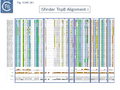
Fig. IS200.34. TnpB Alignment
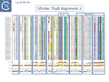
Fig. IS200.34. TnpB Alignment
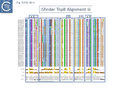
Fig. IS200.34. TnpB Alignment


The relationship between Cas12 and TnpB has strong support from structural modelling [70]: for example Un1Cas12f1 (Cas14a) from an uncultured archeon [76], which functions as an asymmetric dimer and represents a minimal domain organization of the Cas12 group [70]. However, TnpB from ISDra2 (see below) appears to be a monomer [70].

Evolution of TnpB and IscB from an Ancestral RuvC.
In view of the relationship between TnpB, IscB, RuvC and the Cas proteins, the important question of the evolutionary trajectory of these proteins arises. Using various analytic tools, it was concluded that all Cas9 examples identified to date are probably descended from a single IscB derivative ancestor [66]. This contention arose from the observation that the CRISPR-associated IscB derivatives do not form a single clade but are distributed over the IscB phylogenetic tree suggesting that they evolved independently from a single acquisition [66]. Additional IscB derivatives were also identified in this study which led to an evolutionary scenario involving successive acquisition of domains by an ancestral RuvC (Fig. IS200.36). The additional species included a shorter derivative, IsrB, which carried the bridging helix but not the HNH domain and a longer derivative which had acquired a so-called REC domain [66].
TnpB appears to have followed an alternative evolutionary route towards Cas12. In addition, it is thought that TnpB was an ancestor of the eukaryotic Fanzor proteins [77] associated with diverse eukaryotic potential transposable elements.

Functional analysis of TnpB and IscB
Clearly, the relationship between TnpB and IscB and Cas12 and Cas9 respectively suggested that TnpB and IscB might function as RNA guided nucleases which may, in some way, be involved in transposition [70][66] and this has been extensively tested.
TnpB functions as an RNA-guided Endonuclease.
For TnpB, Karvelis, et al.[70] used ISDra2 as a model system. This has the advantage that its transposition behavior has been well characterised [63][64].
In ISDra2, the 3’ end of the upstream tnpA gene overlaps the 5’ end of tnpB. The authors were unable to efficiently express TnpB as a fusion protein but observed that its yield was significantly increased when in its natural context but in which TnpA had been inactivated by mutation. Although the nature of the mutation is not specified in the article, its behavior could be explained if it were an in-frame deletion or other mutation which does not affect C-terminal translation. Since, (Fig. IS200.37) it seems likely that expression of TnpB involves translational coupling [78][79] with TnpA.

TnpB was found to purify with RNA of approximately 150 nts derived from the IS RE (reRNA). reRNA was complementary to the tnpB 3’ end, RE and about 16 nt of (host) flanking DNA (Fig. IS200.38). This RNA with the secondary structure provided by the RE sequence and the 3’ extended flanking DNA is of the expected configuration for relatives of Cas12 (Fig. IS200.36). Previous studies had identified non coding RNA (ncRNA) from the 3’ end of IS1341, a related IS from Halobacterium salinarum NRC-1, called sense overlapping transcripts (sotRNAs) [80].

Such sotRNA transcripts, specific for tnpB genes, had previously been identified by Gomes-Filho et al., [80] in a number of Archaea and Bacteria including S. acidocaldarius, Methanopyrus kandleri, Helicobacter pylori and E. coli K12. There has also been some indication of “transposase-related” sense overlapping transcripts of tnpB-like genes from T. kodakarensis [81] and P. furiosus, [82]. However, that these may represent guide RNAs had not been explicitly considered.
Karvelis et al.[70] demonstrated that TnpB, purified using a His tag, could cleave DNA. They argued that since the 3’ end of the ISDra2 reRNA corresponds to the DNA target, it would vary according to the position of the IS insertion and the reRNA may (have) serve(d) as a guide RNA. If true, cleavage of the target DNA should occur within the 3’ extension sequence of the flank (the foot of RE in Fig. IS200.37). In this context, it is interesting that the (DNA) structure of the right end was shown to form a base triple which is a characteristic of RNA [10].
To determine whether RNA-guided cleavage occurred , they constructed a system (Fig. IS200.38. A) using a plasmid supplying TnpB together with an reRNA which included a 16 (or 20) defined flank sequence and was terminated by a specific Hepatitis delta virus ribozyme (HDV; [83]) to produce a defined 3’ RNA end [84]. A lysate from the host strain was then used in cleavage assays of a library of target plasmids each containing a specific defined 16 base pair sequence directly downstream from a 7 bp (7N) randomised sequence (Fig. IS200.38. B). This has previously been used to identify conserved PAM sequences [70] [85]. Specific double strand cleavage products were captured by adapter ligation (details in Karvelis et al.[70] and the sequence of the resulting enriched 7N region was determined. This corresponded to the conserved ISDra2 target pentanucleotide TTGAT (with a higher enrichment for GA) sequence which is essential for IS insertion and abuts LE in the integrated IS. By equivalence to PAM, this sequence was called TAM (Transposon Adjacent Motif) [70] see also [66].
This cleavage specificity was confirmed using purified TnpB-RNP in which the protein and RNA components were produced by separate plasmids and a target plasmid carrying a 3’ flank and a 5’ TTGAT TAM pentanucleotide and a different guide sequence (Fig. IS200.38.C). The results showed a majority of double strand breaks in the supercoiled target plasmid to generate linear plasmids but also a significant level of nicked product. The TnpB-RNP was also active on a linear substrate. In both cases, use of a TnpB D191A mutant, part of the conserved RuvC DED catalytic triad, eliminated the reaction. Robust TnpB-mediated cleavage activity was observed and required both TAM and guide RNA sequences. Further sequence analysis revealed that cleavage occurred distal to the TAM sequence at the guide sequence boundary and was specific for cleavage on the bottom strand but showed some variation on the top strand (Fig. IS200.38). There are some differences however with Cas12. TnpB is a monomer and requires a single copy of reRNA [70].

Moreover, in vivo, TnpB expression together with reRNA from one plasmid resulted in loss of a second plasmid carrying the reDNA target (interference), presumably as a result of cleavage at the target site and linearisation of the plasmid. This of course may explain the inhibitory effect of TnpB originally observed by Pasternak et al. [64].
Additionally, the authors were able to demonstrate that the system functions in eukaryotic cells opening the possibility that it could be suitably modified for gene editing.
A similar study by Altae-Tran et al.[66] using purified TnpB from a less well characterised tnpB gene of Alicyclobacillus macrosporangiidus, showed that the protein catalysed cleavage of both double- and single-stranded DNA targets in both a TAM-dependent and TAM independent manner. As in the case of TnpBISDra2, A. macrosporangiidus TnpB-associated guide RNA was identified and derived from the 3’ end of the tnpB gene. In this case, the TAM appeared to be the tetranucleotide TCAC.
IscB functions as an RNA-guided Endonuclease
Altae-Tran et al.[66] also examined a very large number of rather disperse IscB systems for their endonuclease properties, their association with RNA and their capacity as RNA guide proteins. Initial studies concerned a CRISPR associated IscB (marked in the article as Delaware Bay acquatic sample), which when purified from a heterologous Escherichia coli host was associated with an RNA localised directly upstream of iscB which generated a signal in a PAM “discovery” assay and was able to generate cleavage products in vitro with the appropriate target.
An alignment of over 500 (non-redundant) iscB genes revealed an upstream region of conserved sequence of about 300 bp which terminated at what the authors state is an IS200/IS605-like end. One specific example examined, present in the host K. racemifer genome in nearly 50 copies, was associated with non-coding RNA species in most cases, which they called ΩRNA, with significant secondary structure potential. An example of K. racemifer IscB was investigated in vitro using a plasmid substrate and shown to: use a target adjacent pentanucleotide TAM, ATAAA; and that cleavage was reprogrammable by changing the complementary RNA extension (guide).
To further characterise IscB, the TAM sequences of 57 examples from a collection of 86 genes from a phylogenetically diverse set of bacteria could be determined; of those 57, 5 were reconstituted with their wRNA and found to active in target cleavage; and one, AwaIscB from Allochromatium warmingii, was chosen for further study.
Biochemically, AwaIscB could cleave double strand DNA in a magnesium dependent reprogrammable way with a temperature optimum of 35-40°C and with RNA guide lengths of between 15 and 45 nts. A mutation of the RuvC E residue eliminated cleavage of the non-target strand while mutation of an H residues in the HNH motif eliminated cleavage of the target strand (as expected for a Cas9-related enzyme; Fig. IS200.31). Mutation of both residues eliminated cleavage altogether. Also like Cas9, cleavage was: TAM (PAM) proximal (3 nts from TAM for the target and 8 or 12 nts for the non-target strands; that the RNP protected DNA from ExoIII digestion 19 nts upstream of the TAM on the target and 6 downstream on the non-target (Fig. IS200.39); and that truncation of the newly identified N-terminal PLMP domain (Fig. Tn200.40 top) eliminated activity.

A Model for TnpB Function During Transposition
The role of these RNA-guided endonucleases remains unknown. It has been suggested that they may mediate RNA-guided TnpA-catalysed transposition although it has been shown that TnpA together with the transposon ends contain all the information for integration into single stranded DNA (references in Mobile DNA paper) and TnpB is not required for transposition. Another suggestion is that they may represent a toxin-type system [66].
While there is considerable information concerning the IS200/IS605 family, very little is known about the mobility of structures which include IscB. Although there is circumstantial evidence that IS1341 related elements are mobile, there is no formal evidence that IscB-containing structures are mobile. The rarity of full length ISC has hampered functional studies.
However, one of the important remaining questions concerning Tn200/Tn605 transposition pathway was the way in which these IS increase their copy number following peeling off the donor daughter “chromatid” to leave a transposon-less donor joint. In the model shown in Fig. IS200.16., excision of a single stranded circular IS intermediate from the lagging strand leaves one double strand copy of the IS in the donor replicon carried by one of the daughter “chromatids” (on the leading strand) and a “donor joint” on the other. In this scenario, no increase in the IS copy number would occur.
The results of Karvelis et al.[70] with ISDra2 provide a solution for this problem. The authors propose a model in which, following excision of the single strand circular IS copy and formation of the donor joint (Fig. IS200.40 A), reRNA/TnpB targeted cleavage of the donor joint is used to initiate a copy/choice replacement of the IS from the remaining IS on the daughter chromatid (Fig. IS200.40 A). This would permit maintenance of donor replicon integrity while assuring an increase in IS copy number via transposition. They point out that conceptually, this in some ways resembles group I intron behavior [86].
TnpA-mediated transposition of the IS200/IS605 family is well documented. The transposition model it is based on in vitro experiments using single-strand oligonucleotides, results from in vivo experiments which implicate DnaG and the observation that the orientation of insertion is correlated with the direction of replication of the target chromosome (see [87] and references therein). The proposed function of TnpB in this process neatly completes the transposition model by offering an explanation of how IS copy number is maintained by replacement of the excised IS at the resulting donor joint. However, it is puzzling that IS1341-like elements of both the TnpB and IscB type have proliferated extensively compared to the “full length” IS. This raises the question of the way in which these genetic objects arose and their function in the cell.
Future studies will be necessary to determine whether they are able to transpose and/or recombine with or without a TnpA-like transposase.
Y1 transposase domestication
There are many examples of eukaryotic transposases whose activities have been appropriated to perform various cellular functions (see [88][89][90]. However, the very few examples of this domestication for prokaryotic enzymes concern Y1 transposases.
TnpAREP and REP/BIME
Recently, a new clade of Y1 transposases (TnpAREP) was found associated with REP/BIME sequences in structures called REPtrons [91][92] (Fig. IS200.7 A). In spite of their compact size, bacterial genomes carry many repetitive sequences, often important for genome function and evolution. Among them, Repetitive Extragenic Palindromic sequences (or REPs) are short DNA repeats of 20-40 bp that can form stem-loop structures preceded by a conserved tetranucleotide (GTAG or GGAG) (Fig. IS200.41). REPs are found in intergenic regions in many bacterial species, particularly in proteobacteria, at high copy number [91][93][94]. There are nearly 590 copies in Escherichia coli K12[95] (Fig. IS200.42) and up to 2200 copies in Pseudomonas sp GM79[94]. REPs can exist as individual units but can cluster in more complex structures called Bacterial Interspersed Mosaic Elements (BIME). These are composed of two individual REPs in inverse orientation (REP and iREP) separated by a short linker of variable length. BIME are often found in consecutive tandem copies (Fig. IS200.41). Several roles have been attributed to these sequences including genome structuring, post-transcriptional regulation and genome plasticity. REPs are known to interact with protein partners such as Integration Host Factor[96], DNA gyrase[97] and DNA polymerase I[98]. REPs also increase mRNA stability and can act as transcriptional terminators[99][100] or as targets for different IS [1][101]. It has also been suggested that REP sequences are involved in REP sequences can downregulate translation of upstream genes dependent on trans-translation. This occurs only if they are within 15 nt of a termination codon. It has been suggested that that REPs can stall ribosomes, leading to mRNA cleavage and induction of the trans-translation process[102]. Recombination at REP sequences has also been shown to be involved in the formation of F’ plasmid derivatives (the classic F plasmid carrying various portions of the chromosome (Fig. IS200.43) from Hfr strains[103]. However, the origin of REPs and their dissemination mechanisms are poorly understood.


Although more complex, REPtrons are reminiscent of IS200 group members (Fig. IS200.41). However REPtrons do not appear to be mobile and, in general, a single copy of a given REPtron co-exists with numerous corresponding REP/BIME and genomes may harbor several distinct REPtrons[91][94]. It has therefore been suggested that REP/BIMEs represent a special type of non-autonomous transposable element mobilizable by TnpAREP.
In vitro analysis of REPtrons: Analysis of E. coli REPtron activity in vitro has shown that, like TnpAIS200/IS605, TnpAREP strictly requires single stranded REP/BIME DNA substrates and is strand specific, only REP can be processed, whereas iREP are refractory to cleavage [92]. Purified E. coli TnpAREP promotes ssREP cleavage (in the linker sequences either 3’ or 5’ to the REP structure) and rejoining, and this activity requires the conserved tetranucleotide GTAG and the bulge in the middle of the REP stem [92][104]. Cleavage in vitro is less specific than that of TnpAIS200/IS605 and occurs at a CT dinucleotide.
In contrast to TnpAIS608 and TnpAISDra2, E. coli TnpAREP is a monomer in solution and in the crystal structure[104]. Moreover, in the co-crystal structure, the short C-terminal tail is inserted into the active site blocking access to an ssDNA. It may, therefore, play a regulatory role in the activity. Indeed C-terminal truncation of TnpAREP resulted in increased cleavage activity relative to the full-length protein in vitro. The biochemical and structural analysis suggested that the GTAG 5’ to the foot of the REP hairpin may play a similar role to the guide sequences GL/R in IS200/IS605. Moreover, structural data also highlighted numerous specific contacts between TnpAREP and GTAG, explaining its importance in the activity and clearly distinguishing TnpAREP from TnpAIS200/IS605, which do not directly contact the guide sequences (Cleavage site recognition). The way by which TnpAREP promotes REP/BIME proliferation through their host genomes remains to be determined.
IStrons
Another role for Y1 transposases was suggested by the identification of chimeric genetic elements widely distributed in the genome of Clostridium difficile[105] and Bacillus cereus group[106][107][108]: IStrons. These combine functional and structural properties of group I introns at their 5’-end with those of an IS element at their 3’-end (Fig. IS200.44). This 3' part contains an IS200/IS605 related sequence including two full length or truncated orfs, tnpA and tnpB, very similar to those found in ISDra2 (D. radiodurans) and ISCpe2 (C. perfringens). IStrons are present at several loci in the same genome, indicating that this element is mobile and may move as a complete genetic unit. All IStron copies analyzed so far are inserted 3’ to the pentanucleotide TTGAT. In vivo, all variants can be efficiently and precisely excised signifying that components necessary for ribozyme activity are present [105]. Little is known about IStron behavior but the data suggest that IS components could mediate the spread of IStron while the intron component could assure splicing.

In vitro oligonucleotide-based assays using purified IStron transposase confirmed that at the DNA level, TTGAT is the LE cleavage site in excision and the target site respectively (Caumont-Sarcos, unpublished). At the RNA level, the same sequence is probably required in the splicing reaction[109]. This would represent a novel type of intron invasion and transposition mechanism and provide a direct link between RNA and DNA worlds.
It is interesting to note that related IStrons have recently been identified which include components of the IS607 family [108][110]. These are characterised by a serine transposase together with a tnpB gene [27].
Bibliography
- ↑ 1.0 1.1 1.2 1.3 Siguier P, Gourbeyre E, Varani A, Ton-Hoang B, Chandler M . Everyman's Guide to Bacterial Insertion Sequences. - Microbiol Spectr: 2015 Apr, 3(2);MDNA3-0030-2014 [PubMed:26104715] [DOI]
- ↑ Thomas J, Pritham EJ . Helitrons, the Eukaryotic Rolling-circle Transposable Elements. - Microbiol Spectr: 2015 Aug, 3(4); [PubMed:26350323] [DOI]
- ↑ 3.0 3.1 Chandler M, de la Cruz F, Dyda F, Hickman AB, Moncalian G, Ton-Hoang B . Breaking and joining single-stranded DNA: the HUH endonuclease superfamily. - Nat Rev Microbiol: 2013 Aug, 11(8);525-38 [PubMed:23832240] [DOI]
- ↑ 4.0 4.1 4.2 4.3 Lam S, Roth JR . IS200: a Salmonella-specific insertion sequence. - Cell: 1983 Oct, 34(3);951-60 [PubMed:6313217] [DOI]
- ↑ 5.0 5.1 5.2 Kersulyte D, Akopyants NS, Clifton SW, Roe BA, Berg DE . Novel sequence organization and insertion specificity of IS605 and IS606: chimaeric transposable elements of Helicobacter pylori. - Gene: 1998 Nov 26, 223(1-2);175-86 [PubMed:9858724] [DOI]
- ↑ Höök-Nikanne J, Berg DE, Peek RM Jr, Kersulyte D, Tummuru MK, Blaser MJ . DNA sequence conservation and diversity in transposable element IS605 of Helicobacter pylori. - Helicobacter: 1998 Jun, 3(2);79-85 [PubMed:9631304] [DOI]
- ↑ 7.0 7.1 7.2 7.3 7.4 7.5 Kersulyte D, Velapatiño B, Dailide G, Mukhopadhyay AK, Ito Y, Cahuayme L, Parkinson AJ, Gilman RH, Berg DE . Transposable element ISHp608 of Helicobacter pylori: nonrandom geographic distribution, functional organization, and insertion specificity. - J Bacteriol: 2002 Feb, 184(4);992-1002 [PubMed:11807059] [DOI]
- ↑ 8.0 8.1 8.2 8.3 8.4 8.5 8.6 Ronning DR, Guynet C, Ton-Hoang B, Perez ZN, Ghirlando R, Chandler M, Dyda F . Active site sharing and subterminal hairpin recognition in a new class of DNA transposases. - Mol Cell: 2005 Oct 7, 20(1);143-54 [PubMed:16209952] [DOI]
- ↑ 9.0 9.1 9.2 9.3 9.4 9.5 9.6 9.7 Ton-Hoang B, Guynet C, Ronning DR, Cointin-Marty B, Dyda F, Chandler M . Transposition of ISHp608, member of an unusual family of bacterial insertion sequences. - EMBO J: 2005 Sep 21, 24(18);3325-38 [PubMed:16163392] [DOI]
- ↑ 10.0 10.1 10.2 10.3 10.4 Guynet C, Hickman AB, Barabas O, Dyda F, Chandler M, Ton-Hoang B . In vitro reconstitution of a single-stranded transposition mechanism of IS608. - Mol Cell: 2008 Feb 15, 29(3);302-12 [PubMed:18280236] [DOI]
- ↑ 11.0 11.1 11.2 11.3 11.4 11.5 11.6 11.7 Barabas O, Ronning DR, Guynet C, Hickman AB, Ton-Hoang B, Chandler M, Dyda F . Mechanism of IS200/IS605 family DNA transposases: activation and transposon-directed target site selection. - Cell: 2008 Jan 25, 132(2);208-20 [PubMed:18243097] [DOI]
- ↑ Pasternak C, Ton-Hoang B, Coste G, Bailone A, Chandler M, Sommer S . Irradiation-induced Deinococcus radiodurans genome fragmentation triggers transposition of a single resident insertion sequence. - PLoS Genet: 2010 Jan 15, 6(1);e1000799 [PubMed:20090938] [DOI]
- ↑ 13.0 13.1 13.2 13.3 13.4 13.5 13.6 Ton-Hoang B, Pasternak C, Siguier P, Guynet C, Hickman AB, Dyda F, Sommer S, Chandler M . Single-stranded DNA transposition is coupled to host replication. - Cell: 2010 Aug 6, 142(3);398-408 [PubMed:20691900] [DOI]
- ↑ 14.0 14.1 14.2 14.3 14.4 14.5 14.6 14.7 Hickman AB, James JA, Barabas O, Pasternak C, Ton-Hoang B, Chandler M, Sommer S, Dyda F . DNA recognition and the precleavage state during single-stranded DNA transposition in D. radiodurans. - EMBO J: 2010 Nov 17, 29(22);3840-52 [PubMed:20890269] [DOI]
- ↑ 15.0 15.1 Filée J, Siguier P, Chandler M . Insertion sequence diversity in archaea. - Microbiol Mol Biol Rev: 2007 Mar, 71(1);121-57 [PubMed:17347521] [DOI]
- ↑ Devalckenaere A, Odaert M, Trieu-Cuot P, Simonet M . Characterization of IS1541-like elements in Yersinia enterocolitica and Yersinia pseudotuberculosis. - FEMS Microbiol Lett: 1999 Jul 1, 176(1);229-33 [PubMed:10418150] [DOI]
- ↑ 17.0 17.1 17.2 Beuzón CR, Chessa D, Casadesús J . IS200: an old and still bacterial transposon. - Int Microbiol: 2004 Mar, 7(1);3-12 [PubMed:15179601]
- ↑ Bisercić M, Ochman H . The ancestry of insertion sequences common to Escherichia coli and Salmonella typhimurium. - J Bacteriol: 1993 Dec, 175(24);7863-8 [PubMed:8253675] [DOI]
- ↑ Bisercić M, Ochman H . Natural populations of Escherichia coli and Salmonella typhimurium harbor the same classes of insertion sequences. - Genetics: 1993 Mar, 133(3);449-54 [PubMed:8384142] [DOI]
- ↑ Beuzón CR, Casadesús J . Conserved structure of IS200 elements in Salmonella. - Nucleic Acids Res: 1997 Apr 1, 25(7);1355-61 [PubMed:9060429] [DOI]
- ↑ 21.0 21.1 Lam S, Roth JR . Structural and functional studies of insertion element IS200. - J Mol Biol: 1986 Jan 20, 187(2);157-67 [PubMed:3009825] [DOI]
- ↑ 22.0 22.1 Beuzón CR, Marqués S, Casadesús J . Repression of IS200 transposase synthesis by RNA secondary structures. - Nucleic Acids Res: 1999 Sep 15, 27(18);3690-5 [PubMed:10471738] [DOI]
- ↑ Sittka A, Lucchini S, Papenfort K, Sharma CM, Rolle K, Binnewies TT, Hinton JC, Vogel J . Deep sequencing analysis of small noncoding RNA and mRNA targets of the global post-transcriptional regulator, Hfq. - PLoS Genet: 2008 Aug 22, 4(8);e1000163 [PubMed:18725932] [DOI]
- ↑ Ellis MJ, Trussler RS, Haniford DB . A cis-encoded sRNA, Hfq and mRNA secondary structure act independently to suppress IS200 transposition. - Nucleic Acids Res: 2015 Jul 27, 43(13);6511-27 [PubMed:26044710] [DOI]
- ↑ Lam S, Roth JR . Genetic mapping of IS200 copies in Salmonella typhimurim strain LT2. - Genetics: 1983 Dec, 105(4);801-11 [PubMed:6315530] [DOI]
- ↑ Odaert M, Devalckenaere A, Trieu-Cuot P, Simonet M . Molecular characterization of IS1541 insertions in the genome of Yersinia pestis. - J Bacteriol: 1998 Jan, 180(1);178-81 [PubMed:9422611] [DOI]
- ↑ 27.0 27.1 Kersulyte D, Mukhopadhyay AK, Shirai M, Nakazawa T, Berg DE . Functional organization and insertion specificity of IS607, a chimeric element of Helicobacter pylori. - J Bacteriol: 2000 Oct, 182(19);5300-8 [PubMed:10986230] [DOI]
- ↑ Boocock MR, Rice PA . A proposed mechanism for IS607-family serine transposases. - Mob DNA: 2013 Nov 6, 4(1);24 [PubMed:24195768] [DOI]
- ↑ Akopyants NS, Fradkov A, Diatchenko L, Hill JE, Siebert PD, Lukyanov SA, Sverdlov ED, Berg DE . PCR-based subtractive hybridization and differences in gene content among strains of Helicobacter pylori. - Proc Natl Acad Sci U S A: 1998 Oct 27, 95(22);13108-13 [PubMed:9789049] [DOI]
- ↑ Islam SM, Hua Y, Ohba H, Satoh K, Kikuchi M, Yanagisawa T, Narumi I . Characterization and distribution of IS8301 in the radioresistant bacterium Deinococcus radiodurans. - Genes Genet Syst: 2003 Oct, 78(5);319-27 [PubMed:14676423] [DOI]
- ↑ 31.0 31.1 Zahradka K, Slade D, Bailone A, Sommer S, Averbeck D, Petranovic M, Lindner AB, Radman M . Reassembly of shattered chromosomes in Deinococcus radiodurans. - Nature: 2006 Oct 5, 443(7111);569-73 [PubMed:17006450] [DOI]
- ↑ Murai N, Kamata H, Nagashima Y, Yagisawa H, Hirata H . A novel insertion sequence (IS)-like element of the thermophilic bacterium PS3 promotes expression of the alanine carrier protein-encoding gene. - Gene: 1995 Sep 22, 163(1);103-7 [PubMed:7557457] [DOI]
- ↑ 33.0 33.1 Bancroft I, Wolk CP . Characterization of an insertion sequence (IS891) of novel structure from the cyanobacterium Anabaena sp. strain M-131. - J Bacteriol: 1989 Nov, 171(11);5949-54 [PubMed:2553665] [DOI]
- ↑ Stanley TL, Ellermeier CD, Slauch JM . Tissue-specific gene expression identifies a gene in the lysogenic phage Gifsy-1 that affects Salmonella enterica serovar typhimurium survival in Peyer's patches. - J Bacteriol: 2000 Aug, 182(16);4406-13 [PubMed:10913072] [DOI]
- ↑ Pasternak C, Ton-Hoang B, Coste G, Bailone A, Chandler M, Sommer S . Irradiation-induced Deinococcus radiodurans genome fragmentation triggers transposition of a single resident insertion sequence. - PLoS Genet: 2010 Jan 15, 6(1);e1000799 [PubMed:20090938] [DOI]
- ↑ Koonin EV, Ilyina TV . Computer-assisted dissection of rolling circle DNA replication. - Biosystems: 1993, 30(1-3);241-68 [PubMed:8374079] [DOI]
- ↑ 37.0 37.1 Lee HH, Yoon JY, Kim HS, Kang JY, Kim KH, Kim DJ, Ha JY, Mikami B, Yoon HJ, Suh SW . Crystal structure of a metal ion-bound IS200 transposase. - J Biol Chem: 2006 Feb 17, 281(7);4261-6 [PubMed:16340015] [DOI]
- ↑ 38.0 38.1 38.2 38.3 38.4 He S, Guynet C, Siguier P, Hickman AB, Dyda F, Chandler M, Ton-Hoang B . IS200/IS605 family single-strand transposition: mechanism of IS608 strand transfer. - Nucleic Acids Res: 2013 Mar 1, 41(5);3302-13 [PubMed:23345619] [DOI]
- ↑ 39.0 39.1 39.2 39.3 39.4 39.5 39.6 39.7 He S, Hickman AB, Dyda F, Johnson NP, Chandler M, Ton-Hoang B . Reconstitution of a functional IS608 single-strand transpososome: role of non-canonical base pairing. - Nucleic Acids Res: 2011 Oct, 39(19);8503-12 [PubMed:21745812] [DOI]
- ↑ 40.0 40.1 Guynet C, Achard A, Hoang BT, Barabas O, Hickman AB, Dyda F, Chandler M . Resetting the site: redirecting integration of an insertion sequence in a predictable way. - Mol Cell: 2009 Jun 12, 34(5);612-9 [PubMed:19524540] [DOI]
- ↑ Morero NR, Zuliani C, Kumar B, Bebel A, Okamoto S, Guynet C, Hickman AB, Chandler M, Dyda F, Barabas O . Targeting IS608 transposon integration to highly specific sequences by structure-based transposon engineering. - Nucleic Acids Res: 2018 May 4, 46(8);4152-4163 [PubMed:29635476] [DOI]
- ↑ Pasternak C, Ton-Hoang B, Coste G, Bailone A, Chandler M, Sommer S . Irradiation-induced Deinococcus radiodurans genome fragmentation triggers transposition of a single resident insertion sequence. - PLoS Genet: 2010 Jan 15, 6(1);e1000799 [PubMed:20090938] [DOI]
- ↑ 43.0 43.1 Mennecier S, Servant P, Coste G, Bailone A, Sommer S . Mutagenesis via IS transposition in Deinococcus radiodurans. - Mol Microbiol: 2006 Jan, 59(1);317-25 [PubMed:16359337] [DOI]
- ↑ Parks AR, Li Z, Shi Q, Owens RM, Jin MM, Peters JE . Transposition into replicating DNA occurs through interaction with the processivity factor. - Cell: 2009 Aug 21, 138(4);685-95 [PubMed:19703395] [DOI]
- ↑ Hu WY, Derbyshire KM . Target choice and orientation preference of the insertion sequence IS903. - J Bacteriol: 1998 Jun, 180(12);3039-48 [PubMed:9620951] [DOI]
- ↑ Roberts D, Hoopes BC, McClure WR, Kleckner N . IS10 transposition is regulated by DNA adenine methylation. - Cell: 1985 Nov, 43(1);117-30 [PubMed:3000598] [DOI]
- ↑ Yin JC, Krebs MP, Reznikoff WS . Effect of dam methylation on Tn5 transposition. - J Mol Biol: 1988 Jan 5, 199(1);35-45 [PubMed:2451025] [DOI]
- ↑ Dodson KW, Berg DE . Factors affecting transposition activity of IS50 and Tn5 ends. - Gene: 1989, 76(2);207-13 [PubMed:2546858] [DOI]
- ↑ Spradling AC, Bellen HJ, Hoskins RA . Drosophila P elements preferentially transpose to replication origins. - Proc Natl Acad Sci U S A: 2011 Sep 20, 108(38);15948-53 [PubMed:21896744] [DOI]
- ↑ Zechner EL, Wu CA, Marians KJ . Coordinated leading- and lagging-strand synthesis at the Escherichia coli DNA replication fork. III. A polymerase-primase interaction governs primer size. - J Biol Chem: 1992 Feb 25, 267(6);4054-63 [PubMed:1531480]
- ↑ Wu CA, Zechner EL, Reems JA, McHenry CS, Marians KJ . Coordinated leading- and lagging-strand synthesis at the Escherichia coli DNA replication fork. V. Primase action regulates the cycle of Okazaki fragment synthesis. - J Biol Chem: 1992 Feb 25, 267(6);4074-83 [PubMed:1740453]
- ↑ Lau IF, Filipe SR, Søballe B, Økstad OA, Barre FX, Sherratt DJ . Spatial and temporal organization of replicating Escherichia coli chromosomes. - Mol Microbiol: 2003 Aug, 49(3);731-43 [PubMed:12864855] [DOI]
- ↑ 53.0 53.1 53.2 Lavatine L, He S, Caumont-Sarcos A, Guynet C, Marty B, Chandler M, Ton-Hoang B . Single strand transposition at the host replication fork. - Nucleic Acids Res: 2016 Sep 19, 44(16);7866-83 [PubMed:27466393] [DOI]
- ↑ Hansen MT . Multiplicity of genome equivalents in the radiation-resistant bacterium Micrococcus radiodurans. - J Bacteriol: 1978 Apr, 134(1);71-5 [PubMed:649572] [DOI]
- ↑ Harsojo, Kitayama S, Matsuyama A . Genome multiplicity and radiation resistance in Micrococcus radiodurans. - J Biochem: 1981 Sep, 90(3);877-80 [PubMed:7309705] [DOI]
- ↑ Pasternak C, Ton-Hoang B, Coste G, Bailone A, Chandler M, Sommer S . Irradiation-induced Deinococcus radiodurans genome fragmentation triggers transposition of a single resident insertion sequence. - PLoS Genet: 2010 Jan 15, 6(1);e1000799 [PubMed:20090938] [DOI]
- ↑ Pasternak C, Ton-Hoang B, Coste G, Bailone A, Chandler M, Sommer S . Irradiation-induced Deinococcus radiodurans genome fragmentation triggers transposition of a single resident insertion sequence. - PLoS Genet: 2010 Jan 15, 6(1);e1000799 [PubMed:20090938] [DOI]
- ↑ Kim NH, Lee G, Sherer NA, Martini KM, Goldenfeld N, Kuhlman TE . Real-time transposable element activity in individual live cells. - Proc Natl Acad Sci U S A: 2016 Jun 28, 113(26);7278-83 [PubMed:27298350] [DOI]
- ↑ Markwardt ML, Kremers GJ, Kraft CA, Ray K, Cranfill PJ, Wilson KA, Day RN, Wachter RM, Davidson MW, Rizzo MA . An improved cerulean fluorescent protein with enhanced brightness and reduced reversible photoswitching. - PLoS One: 2011 Mar 29, 6(3);e17896 [PubMed:21479270] [DOI]
- ↑ Nagai T, Ibata K, Park ES, Kubota M, Mikoshiba K, Miyawaki A . A variant of yellow fluorescent protein with fast and efficient maturation for cell-biological applications. - Nat Biotechnol: 2002 Jan, 20(1);87-90 [PubMed:11753368] [DOI]
- ↑ Shapiro JA, Higgins NP . Variation of beta-galactosidase expression from Mudlac elements during the development of Escherichia coli colonies. - Ann Inst Pasteur Microbiol: 1988 Jan-Feb, 139(1);79-103 [PubMed:2838063] [DOI]
- ↑ Shapiro JA, Higgins NP . Differential activity of a transposable element in Escherichia coli colonies. - J Bacteriol: 1989 Nov, 171(11);5975-86 [PubMed:2553666] [DOI]
- ↑ 63.0 63.1 Pasternak C, Ton-Hoang B, Coste G, Bailone A, Chandler M, Sommer S . Irradiation-induced Deinococcus radiodurans genome fragmentation triggers transposition of a single resident insertion sequence. - PLoS Genet: 2010 Jan 15, 6(1);e1000799 [PubMed:20090938] [DOI]
- ↑ 64.0 64.1 64.2 64.3 Pasternak C, Dulermo R, Ton-Hoang B, Debuchy R, Siguier P, Coste G, Chandler M, Sommer S . ISDra2 transposition in Deinococcus radiodurans is downregulated by TnpB. - Mol Microbiol: 2013 Apr, 88(2);443-55 [PubMed:23461641] [DOI]
- ↑ 65.00 65.01 65.02 65.03 65.04 65.05 65.06 65.07 65.08 65.09 65.10 65.11 65.12 65.13 65.14 65.15 65.16
Kapitonov VV, Makarova KS, Koonin EV . ISC, a Novel Group of Bacterial and Archaeal DNA Transposons That Encode Cas9 Homologs. - J Bacteriol: 2015 Dec 28, 198(5);797-807 [PubMed:26712934]
[DOI]
- ↑ 66.00 66.01 66.02 66.03 66.04 66.05 66.06 66.07 66.08 66.09 66.10 66.11 66.12 66.13 66.14 66.15 66.16 Altae-Tran H, Kannan S, Demircioglu FE, Oshiro R, Nety SP, McKay LJ, Dlakić M, Inskeep WP, Makarova KS, Macrae RK, Koonin EV, Zhang F . The widespread IS200/IS605 transposon family encodes diverse programmable RNA-guided endonucleases. - Science: 2021 Oct, 374(6563);57-65 [PubMed:34591643] [DOI]
- ↑ Chylinski K, Makarova KS, Charpentier E, Koonin EV . Classification and evolution of type II CRISPR-Cas systems. - Nucleic Acids Res: 2014 Jun, 42(10);6091-105 [PubMed:24728998] [DOI]
- ↑ Shmakov S, Smargon A, Scott D, Cox D, Pyzocha N, Yan W, Abudayyeh OO, Gootenberg JS, Makarova KS, Wolf YI, Severinov K, Zhang F, Koonin EV . Diversity and evolution of class 2 CRISPR-Cas systems. - Nat Rev Microbiol: 2017 Mar, 15(3);169-182 [PubMed:28111461] [DOI]
- ↑ Makarova KS, Wolf YI, Iranzo J, Shmakov SA, Alkhnbashi OS, Brouns SJJ, Charpentier E, Cheng D, Haft DH, Horvath P, Moineau S, Mojica FJM, Scott D, Shah SA, Siksnys V, Terns MP, Venclovas Č, White MF, Yakunin AF, Yan W, Zhang F, Garrett RA, Backofen R, van der Oost J, Barrangou R, Koonin EV . Evolutionary classification of CRISPR-Cas systems: a burst of class 2 and derived variants. - Nat Rev Microbiol: 2020 Feb, 18(2);67-83 [PubMed:31857715] [DOI]
- ↑ 70.00 70.01 70.02 70.03 70.04 70.05 70.06 70.07 70.08 70.09 70.10 70.11 70.12 70.13 70.14 70.15 70.16 70.17 70.18 70.19 70.20 70.21 70.22 Karvelis T, Druteika G, Bigelyte G, Budre K, Zedaveinyte R, Silanskas A, Kazlauskas D, Venclovas Č, Siksnys V . Transposon-associated TnpB is a programmable RNA-guided DNA endonuclease. - Nature: 2021 Nov, 599(7886);692-696 [PubMed:34619744] [DOI]
- ↑ 71.0 71.1 Jinek M, Jiang F, Taylor DW, Sternberg SH, Kaya E, Ma E, Anders C, Hauer M, Zhou K, Lin S, Kaplan M, Iavarone AT, Charpentier E, Nogales E, Doudna JA . Structures of Cas9 endonucleases reveal RNA-mediated conformational activation. - Science: 2014 Mar 14, 343(6176);1247997 [PubMed:24505130] [DOI]
- ↑ 72.0 72.1 Gasiunas G, Barrangou R, Horvath P, Siksnys V . Cas9-crRNA ribonucleoprotein complex mediates specific DNA cleavage for adaptive immunity in bacteria. - Proc Natl Acad Sci U S A: 2012 Sep 25, 109(39);E2579-86 [PubMed:22949671] [DOI]
- ↑ Makarova KS, Aravind L, Wolf YI, Koonin EV . Unification of Cas protein families and a simple scenario for the origin and evolution of CRISPR-Cas systems. - Biol Direct: 2011 Jul 14, 6;38 [PubMed:21756346] [DOI]
- ↑ Swarts DC . Stirring Up the Type V Alphabet Soup. - CRISPR J: 2019 Feb, 2;14-16 [PubMed:31021231] [DOI]
- ↑ Xiao R, Li Z, Wang S, Han R, Chang L . Structural basis for substrate recognition and cleavage by the dimerization-dependent CRISPR-Cas12f nuclease. - Nucleic Acids Res: 2021 Apr 19, 49(7);4120-4128 [PubMed:33764415] [DOI]
- ↑ Takeda SN, Nakagawa R, Okazaki S, Hirano H, Kobayashi K, Kusakizako T, Nishizawa T, Yamashita K, Nishimasu H, Nureki O . Structure of the miniature type V-F CRISPR-Cas effector enzyme. - Mol Cell: 2021 Feb 4, 81(3);558-570.e3 [PubMed:33333018] [DOI]
- ↑ Bao W, Jurka J . Homologues of bacterial TnpB_IS605 are widespread in diverse eukaryotic transposable elements. - Mob DNA: 2013 Apr 1, 4(1);12 [PubMed:23548000] [DOI]
- ↑ Rex G, Surin B, Besse G, Schneppe B, McCarthy JE . The mechanism of translational coupling in Escherichia coli. Higher order structure in the atpHA mRNA acts as a conformational switch regulating the access of de novo initiating ribosomes. - J Biol Chem: 1994 Jul 8, 269(27);18118-27 [PubMed:7517937]
- ↑
Huber M, Faure G, Laass S, Kolbe E, Seitz K, Wehrheim C, Wolf YI, Koonin EV, Soppa J . Translational coupling via termination-reinitiation in archaea and bacteria. - Nat Commun: 2019 Sep 5, 10(1);4006 [PubMed:31488843]
[DOI]
- ↑ 80.0 80.1 Gomes-Filho JV, Zaramela LS, Italiani VC, Baliga NS, Vêncio RZ, Koide T . Sense overlapping transcripts in IS1341-type transposase genes are functional non-coding RNAs in archaea. - RNA Biol: 2015, 12(5);490-500 [PubMed:25806405] [DOI]
- ↑ Jäger D, Förstner KU, Sharma CM, Santangelo TJ, Reeve JN . Primary transcriptome map of the hyperthermophilic archaeon Thermococcus kodakarensis. - BMC Genomics: 2014 Aug 16, 15(1);684 [PubMed:25127548] [DOI]
- ↑ Klein RJ, Misulovin Z, Eddy SR . Noncoding RNA genes identified in AT-rich hyperthermophiles. - Proc Natl Acad Sci U S A: 2002 May 28, 99(11);7542-7 [PubMed:12032319] [DOI]
- ↑ Been MD, Wickham GS . Self-cleaving ribozymes of hepatitis delta virus RNA. - Eur J Biochem: 1997 Aug 1, 247(3);741-53 [PubMed:9288893] [DOI]
- ↑ Ferré-D'Amaré AR, Zhou K, Doudna JA . Crystal structure of a hepatitis delta virus ribozyme. - Nature: 1998 Oct 8, 395(6702);567-74 [PubMed:9783582] [DOI]
- ↑ Karvelis T, Young JK, Siksnys V . A pipeline for characterization of novel Cas9 orthologs. - Methods Enzymol: 2019, 616;219-240 [PubMed:30691644] [DOI]
- ↑ Stoddard BL . Homing endonucleases from mobile group I introns: discovery to genome engineering. - Mob DNA: 2014 Mar 3, 5(1);7 [PubMed:24589358] [DOI]
- ↑ He S, Corneloup A, Guynet C, Lavatine L, Caumont-Sarcos A, Siguier P, Marty B, Dyda F, Chandler M, Ton Hoang B . The IS200/IS605 Family and "Peel and Paste" Single-strand Transposition Mechanism. - Microbiol Spectr: 2015 Aug, 3(4); [PubMed:26350330] [DOI]
- ↑ Volff JN . Turning junk into gold: domestication of transposable elements and the creation of new genes in eukaryotes. - Bioessays: 2006 Sep, 28(9);913-22 [PubMed:16937363] [DOI]
- ↑ Vogt A, Goldman AD, Mochizuki K, Landweber LF . Transposon domestication versus mutualism in ciliate genome rearrangements. - PLoS Genet: 2013, 9(8);e1003659 [PubMed:23935529] [DOI]
- ↑ Vogt A, Mochizuki K . A domesticated PiggyBac transposase interacts with heterochromatin and catalyzes reproducible DNA elimination in Tetrahymena. - PLoS Genet: 2013, 9(12);e1004032 [PubMed:24348275] [DOI]
- ↑ 91.0 91.1 91.2 Nunvar J, Huckova T, Licha I . Identification and characterization of repetitive extragenic palindromes (REP)-associated tyrosine transposases: implications for REP evolution and dynamics in bacterial genomes. - BMC Genomics: 2010 Jan 19, 11;44 [PubMed:20085626] [DOI]
- ↑ 92.0 92.1 92.2 Ton-Hoang B, Siguier P, Quentin Y, Onillon S, Marty B, Fichant G, Chandler M . Structuring the bacterial genome: Y1-transposases associated with REP-BIME sequences. - Nucleic Acids Res: 2012 Apr, 40(8);3596-609 [PubMed:22199259] [DOI]
- ↑ Rocco F, De Gregorio E, Di Nocera PP . A giant family of short palindromic sequences in Stenotrophomonas maltophilia. - FEMS Microbiol Lett: 2010 Jul, 308(2);185-92 [PubMed:20528935] [DOI]
- ↑ 94.0 94.1 94.2 Nunvar J, Licha I, Schneider B . Evolution of REP diversity: a comparative study. - BMC Genomics: 2013 Jun 10, 14;385 [PubMed:23758774] [DOI]
- ↑ 95.0 95.1 95.2 Gilson E, Bachellier S, Perrin S, Perrin D, Grimont PA, Grimont F, Hofnung M . Palindromic unit highly repetitive DNA sequences exhibit species specificity within Enterobacteriaceae. - Res Microbiol: 1990 Nov-Dec, 141(9);1103-16 [PubMed:2092362] [DOI]
- ↑ Boccard F, Prentki P . Specific interaction of IHF with RIBs, a class of bacterial repetitive DNA elements located at the 3' end of transcription units. - EMBO J: 1993 Dec 15, 12(13);5019-27 [PubMed:8262044] [DOI]
- ↑ Espéli O, Boccard F . In vivo cleavage of Escherichia coli BIME-2 repeats by DNA gyrase: genetic characterization of the target and identification of the cut site. - Mol Microbiol: 1997 Nov, 26(4);767-77 [PubMed:9427406] [DOI]
- ↑ Gilson E, Perrin D, Hofnung M . DNA polymerase I and a protein complex bind specifically to E. coli palindromic unit highly repetitive DNA: implications for bacterial chromosome organization. - Nucleic Acids Res: 1990 Jul 11, 18(13);3941-52 [PubMed:2197600] [DOI]
- ↑ Rocco F, De Gregorio E, Di Nocera PP . A giant family of short palindromic sequences in Stenotrophomonas maltophilia. - FEMS Microbiol Lett: 2010 Jul, 308(2);185-92 [PubMed:20528935] [DOI]
- ↑ Khemici V, Carpousis AJ . The RNA degradosome and poly(A) polymerase of Escherichia coli are required in vivo for the degradation of small mRNA decay intermediates containing REP-stabilizers. - Mol Microbiol: 2004 Feb, 51(3);777-90 [PubMed:14731278] [DOI]
- ↑ Tobes R, Pareja E . Bacterial repetitive extragenic palindromic sequences are DNA targets for Insertion Sequence elements. - BMC Genomics: 2006 Mar 24, 7;62 [PubMed:16563168] [DOI]
- ↑ Liang W, Rudd KE, Deutscher MP . A role for REP sequences in regulating translation. - Mol Cell: 2015 May 7, 58(3);431-9 [PubMed:25891074] [DOI]
- ↑ Kofoid E, Bergthorsson U, Slechta ES, Roth JR . Formation of an F' plasmid by recombination between imperfectly repeated chromosomal Rep sequences: a closer look at an old friend (F'(128) pro lac). - J Bacteriol: 2003 Jan, 185(2);660-3 [PubMed:12511513] [DOI]
- ↑ 104.0 104.1 Messing SA, Ton-Hoang B, Hickman AB, McCubbin AJ, Peaslee GF, Ghirlando R, Chandler M, Dyda F . The processing of repetitive extragenic palindromes: the structure of a repetitive extragenic palindrome bound to its associated nuclease. - Nucleic Acids Res: 2012 Oct, 40(19);9964-79 [PubMed:22885300] [DOI]
- ↑ 105.0 105.1 Braun V, Mehlig M, Moos M, Rupnik M, Kalt B, Mahony DE, von Eichel-Streiber C . A chimeric ribozyme in clostridium difficile combines features of group I introns and insertion elements. - Mol Microbiol: 2000 Jun, 36(6);1447-59 [PubMed:10931294] [DOI]
- ↑ Tourasse NJ, Helgason E, Økstad OA, Hegna IK, Kolstø AB . The Bacillus cereus group: novel aspects of population structure and genome dynamics. - J Appl Microbiol: 2006 Sep, 101(3);579-93 [PubMed:16907808] [DOI]
- ↑ Tourasse NJ, Kolstø AB . Survey of group I and group II introns in 29 sequenced genomes of the Bacillus cereus group: insights into their spread and evolution. - Nucleic Acids Res: 2008 Aug, 36(14);4529-48 [PubMed:18587153] [DOI]
- ↑ 108.0 108.1 Tourasse NJ, Stabell FB, Reiter L, Kolstø AB . Unusual group II introns in bacteria of the Bacillus cereus group. - J Bacteriol: 2005 Aug, 187(15);5437-51 [PubMed:16030238] [DOI]
- ↑ Nielsen H, Johansen SD . Group I introns: Moving in new directions. - RNA Biol: 2009 Sep-Oct, 6(4);375-83 [PubMed:19667762] [DOI]
- ↑ Tourasse NJ, Stabell FB, Kolstø AB . Survey of chimeric IStron elements in bacterial genomes: multiple molecular symbioses between group I intron ribozymes and DNA transposons. - Nucleic Acids Res: 2014 Nov 10, 42(20);12333-51 [PubMed:25324310] [DOI]